Human anatomy is the scientific study of the body’s structures. Some of these structures are very small and can only be observed and analyzed with the assistance of a microscope. Other larger structures can readily be seen, manipulated, measured, and weighed. The word “anatomy” comes from a Greek root that means “to cut apart.” Human anatomy was first studied by observing the exterior of the body and observing the wounds of soldiers and other injuries. Later, physicians were allowed to dissect bodies of the dead to augment their knowledge. When a body is dissected, its structures are cut apart in order to observe their physical attributes and their relationships to one another. Dissection is still used in medical schools, anatomy courses, and in pathology labs. In order to observe structures in living people, however, a number of imaging techniques have been developed. These techniques allow clinicians to visualize structures inside the living body such as a cancerous tumor or a fractured bone.
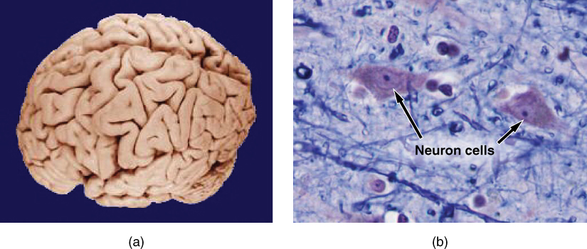
Like most scientific disciplines, anatomy has areas of specialization. Gross anatomy is the study of the larger structures of the body, those visible without the aid of magnification (Figure 1a). Macro- means “large,” thus, gross anatomy is also referred to as macroscopic anatomy. In contrast, micro- means “small,” and microscopic anatomy is the study of structures that can be observed only with the use of a microscope or other magnification devices (Figure 1b). Microscopic anatomy includes cytology, the study of cells and histology, the study of tissues. As the technology of microscopes has advanced, anatomists have been able to observe smaller and smaller structures of the body, from slices of large structures like the heart, to the three-dimensional structures of large molecules in the body.

Figure 1. Gross and Microscopic Anatomy. (a) Gross anatomy considers large structures such as the brain. (b) Microscopic anatomy can deal with the same structures, though at a different scale. This is a micrograph of nerve cells from the brain. LM × 1600. (credit a: “WriterHound”/Wikimedia Commons; credit b: Micrograph provided by the Regents of University of Michigan Medical School © 2012)
Like most scientific disciplines, anatomy has areas of specialization. Gross anatomy is the study of the larger structures of the body, those visible without the aid of magnification (Figure 1a). Macro- means “large,” thus, gross anatomy is also referred to as macroscopic anatomy. In contrast, micro- means “small,” and microscopic anatomy is the study of structures that can be observed only with the use of a microscope or other magnification devices (Figure 1b). Microscopic anatomy includes cytology, the study of cells and histology, the study of tissues. As the technology of microscopes has advanced, anatomists have been able to observe smaller and smaller structures of the body, from slices of large structures like the heart, to the three-dimensional structures of large molecules in the body.

Figure 1. Gross and Microscopic Anatomy. (a) Gross anatomy considers large structures such as the brain. (b) Microscopic anatomy can deal with the same structures, though at a different scale. This is a micrograph of nerve cells from the brain. LM × 1600. (credit a: “WriterHound”/Wikimedia Commons; credit b: Micrograph provided by the Regents of University of Michigan Medical School © 2012)
No comments:
Post a Comment