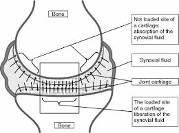
The anatomical parts that suffer the most when there is shortage of water the human body are those without a direct vascular circulation. The anatomical parts that depend for the supply of their needs on the seepage of tissue fluids through another organ suffer most. They will not receive what they need ; the mediating organ will trim its transit routes. The anatomical parts that are excellent examples of this thesis are the joint cartilage (see Fig 28 and Fig 29) and the intervertebral discs. I have explained the mechanisms involved in disc hydration. I will now try to explain the basic problem in the feeding of joint cartilage that is the root cause of damage in the rheumatoid joints and their pain signal, be they the finger joints, the knees joints, or the vertebral joints.
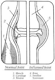

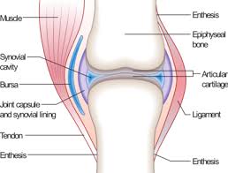
Remember , there is no functioning "dead" part in the body that is nature-designed. All tissues of the human body, including bone, cartilage, and even the disc core are composed of living cells that have to remain alive and be reproductive of daughter cells (except the brain cells that are not replaced before they die) for that particular organ to function. The dead tissues (group of cells) is eaten away by the "garbage collectors" and new tissues replaces them. For the tissue to remain alive, the most simple and initial need is water itself, and then whatever food nutrients supply the water can bring with it.

Fig.28: The normal finger joint demonstrating the common arterial supply to the area. The artery to the capsule can dilate to bring increased blood circulation to the soft tissues of the joint. the artery that goes through the bone canal is restricted by the size of its passageway.
The bone connections of the fingers, hands, and most movement-supporting joints are separated by means of cartilage is connected are thin, whereas the wall of their shaft are made of solid and thicker tube-like bones. The artery of the bone goes through and divides in the canal systems in the thick section of the bone.
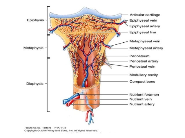
The canals in the bone act as though they are straightjackets (see fig.28) that may not permit dilation of the vessels, even if the vessels themselves could dilate to increase the circulation to the area (the very mechanism that brings greater circulation to the capsule of the joint, whereas the artery that goes through the bone is severely restricted by the fixed size of its canal through the bone). Each bone of the skeleton has only one (very rarely two) artery to feed that bone. Inside the hollow spaces of these bones, nature has housed the manufacturing system of the blood cells ー red cells as well as all the variations of white cells. Nature gives priority to the development of these cells, which entirely depend on many different functions of water in particular. When there is dehydration, there will not be enough water to supply the end bone cartilage with its needs ーFig.29; the blood manufacturing system exercises its priority by means of specially active cation pumps that force the water into the expanding blood cells, which must consist of at least 75 percent water.
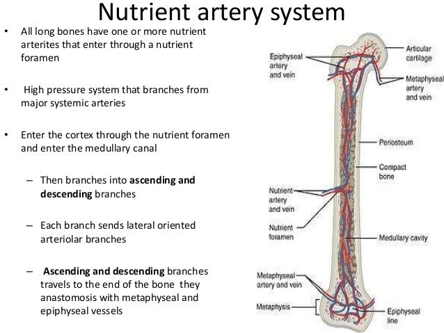
Fig.29
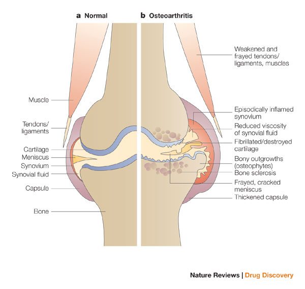
Fig. 29: The left side of the figure represents a normal flow of "water" and nutrient needs of the cartilage covering of the joint bones. The right side represents a decreased flow of "water" to the cartilage covering through the bone. The arterial supply to the capsule becomes increased, causing swelling and effusion into the joint. This route of supply of nutrients is not completely effective for the growing end of the cartilage covering the bones. Forced activity of the joint will damage the cartilage and leave the bone exposed and permanently damaged.

A well-hydrated human body and normal direction of water flow to the joint cartilage:
• The artery to the capsule( normal size).
• The direction of the water flow to the cartilage in a well-hydrate body is towards the bone joint.
• Normal cartilage covering the bone heads of the joint.
• The artery to the capsule (normal size)
A dehydrated human body exposing reduced local flow of water through the bone marrow causing joint cartilage damage:
• Enlarged capsule artery.
• The inflamed and thickened joint cartilage.
• Flow of water and proteins to the joint through the capsule.
• Irregular head of bones after the loss of cartilage.
• Reduced rate of water flow through the bone marrow in a dehydrated body.
Remember , there is no functioning "dead" part in the body that is nature-designed. All tissues of the human body, including bone, cartilage, and even the disc core are composed of living cells that have to remain alive and be reproductive of daughter cells (except the brain cells that are not replaced before they die) for that particular organ to function. The dead tissues (group of cells) is eaten away by the "garbage collectors" and new tissues replaces them. For the tissue to remain alive, the most simple and initial need is water itself, and then whatever food nutrients supply the water can bring with it.
Fig.28: The normal finger joint demonstrating the common arterial supply to the area. The artery to the capsule can dilate to bring increased blood circulation to the soft tissues of the joint. the artery that goes through the bone canal is restricted by the size of its passageway.
The bone connections of the fingers, hands, and most movement-supporting joints are separated by means of cartilage is connected are thin, whereas the wall of their shaft are made of solid and thicker tube-like bones. The artery of the bone goes through and divides in the canal systems in the thick section of the bone.
The canals in the bone act as though they are straightjackets (see fig.28) that may not permit dilation of the vessels, even if the vessels themselves could dilate to increase the circulation to the area (the very mechanism that brings greater circulation to the capsule of the joint, whereas the artery that goes through the bone is severely restricted by the fixed size of its canal through the bone). Each bone of the skeleton has only one (very rarely two) artery to feed that bone. Inside the hollow spaces of these bones, nature has housed the manufacturing system of the blood cells ー red cells as well as all the variations of white cells. Nature gives priority to the development of these cells, which entirely depend on many different functions of water in particular. When there is dehydration, there will not be enough water to supply the end bone cartilage with its needs ーFig.29; the blood manufacturing system exercises its priority by means of specially active cation pumps that force the water into the expanding blood cells, which must consist of at least 75 percent water.
Fig.29
Fig. 29: The left side of the figure represents a normal flow of "water" and nutrient needs of the cartilage covering of the joint bones. The right side represents a decreased flow of "water" to the cartilage covering through the bone. The arterial supply to the capsule becomes increased, causing swelling and effusion into the joint. This route of supply of nutrients is not completely effective for the growing end of the cartilage covering the bones. Forced activity of the joint will damage the cartilage and leave the bone exposed and permanently damaged.
A well-hydrated human body and normal direction of water flow to the joint cartilage:
• The artery to the capsule( normal size).
• The direction of the water flow to the cartilage in a well-hydrate body is towards the bone joint.
• Normal cartilage covering the bone heads of the joint.
• The artery to the capsule (normal size)
A dehydrated human body exposing reduced local flow of water through the bone marrow causing joint cartilage damage:
• Enlarged capsule artery.
• The inflamed and thickened joint cartilage.
• Flow of water and proteins to the joint through the capsule.
• Irregular head of bones after the loss of cartilage.
• Reduced rate of water flow through the bone marrow in a dehydrated body.


