Showing posts with label mouth. Show all posts

Saturday, July 1, 2017

how your digestive system works

How long does it take to digest food — from the time you eat it to the time you excrete it? 
The digestion process takes between 24 and 72 hours, Elimination of undigested residue food  usually begins after 24 hours. Complete elimination from the body may take several days. 

Answers from Michael F. Picco, M.D.
Digestion time varies between individuals and between men and women. After you eat, it takes about six to eight hours for food to pass through your stomach and small intestine. Food then enters your large intestine (colon) for further digestion, absorption of water and, finally, elimination of undigested food.

In the 1980s, Mayo Clinic researchers measured digestion time in 21 healthy people. Total transit time, from eating to elimination in stool, averaged 53 hours (although that figure is a little overstated, because the markers used by the researchers passed more slowly through the stomach than actual food). The average transit time through just the large intestine (colon) was 40 hours, with significant difference between men and women: 33 hours for men, 47 hours for women.

Two British doctors studied digestion time in children. They fed 35 children juice containing a red marker and asked the children's mothers to note when the stool first turned red. The mean time of transit from mouth to anus for the group was 33 hours (meaning half the children had digestion times slower than this and half had digestion times greater than this).

With Regards,
Michael F. Picco, M.D.


Illustration of digestive system

Your digestive system gears up
A fresh-baked apple pie has just come out of the oven. Just the sight and smell of it are enough to make you start salivating. So even before you take a bite, your digestive system has swung into action.

After the first morsel enters your mouth, the many organs of your digestive tract kick into high gear. Here's a look at how your digestive system works, from top to bottom.

Illustration of mouth and salivary glands

Mouth and salivary glands
After you take your first bite of pie, your salivary glands produce saliva — a mixture of secretions that help lubricate and break down food. Besides the salivary glands in the lining of your mouth, you have three pairs of larger salivary glands — the parotid, sublingual and submandibular glands. You typically produce about 2 pints (about 1 liter) of saliva a day.

Not all of the work is chemical, though. As you savor the bite of pie, your teeth work to break down the pie while your tongue mixes it with saliva. This action transforms it into a soft, moist, rounded mass (bolus) suitable for swallowing.

Illustration of esophagus

Esophagus
As you swallow the bite of pie, muscles in your mouth and throat propel it to your upper esophagus, the tube that connects your throat to your stomach. Muscles in the wall of your esophagus create synchronized waves — one after another — that propel the pie into your stomach. In this process, called peristalsis, muscles behind the bolus of pie contract, squeezing it forward, while muscles ahead of it relax, allowing it to advance without resistance.

When the bolus reaches the lower end of your esophagus, pressure from the food signals a muscular valve — the lower esophageal sphincter — to relax and let the food enter your stomach.

Illustration of stomach

Stomach
After entering your stomach, the pie is broken down further. With its powerful muscles, the stomach begins churning and mixing the food into smaller and smaller pieces. Your digestive glands in your stomach lining produce stomach acid and enzymes, which mix with the food to form a murky semifluid or paste called chyme.

Once the chyme is well-mixed, waves of muscle contractions propel it through a valve called the pylorus and into the first section of your small intestine (duodenum). The pylorus might release about an eighth of an ounce (about 4 milliliters) of chyme at a time. The rest is held back for more mixing.

Illustration of pancreas, liver and gallbladder

Pancreas, liver and gallbladder
In your duodenum, digestion continues as chyme from the stomach mixes with a variety of digestive juices from your pancreas, liver and gallbladder:

Pancreas. The pancreas produces digestive enzymes that help break down proteins, carbohydrates and fats.

Liver. The liver produces bile, a solution that helps you digest fats.

Gallbladder. The gallbladder stores bile. As fatty food enters the upper portion of your small intestine (the duodenum), the gallbladder squeezes bile into the small intestine through the bile ducts.

Illustration of small intestine

Small intestine
As bile and pancreatic digestive juices mix with other juices secreted by the wall of your small intestine, digestion continues. What was once apple pie is propelled into the second portion of your small intestine, the jejunum. Here it's further broken down into smaller molecules of nutrients that can be absorbed. Then it moves into the final and longest portion of your small intestine — the ileum — where virtually all of the remaining nutrients are absorbed through the lining of the ileum's wall.

What remains of the food when it reaches the end of the ileum is a combination of water, electrolytes — such as sodium and chloride — and waste products, such as plant fiber and dead cells shed from the lining of your digestive tract.

Illustration of large intestine

Large intestine
As this residue passes through the colon, nearly all of the water is absorbed, leaving a usually soft but formed substance called stool. Muscles in the wall of your colon separate the waste into small segments that are pushed into your lower colon and rectum. As the rectal walls are stretched, they signal the need for a bowel movement.

When the sphincter muscles in your anus relax, the rectal walls contract to increase pressure. These coordinated muscle contractions expel the stool.

The End of Slide Show. 

Monday, April 3, 2017

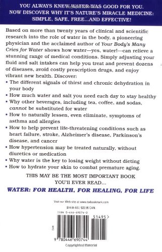
Water: For Health, for Healing, for Life: You're Not Sick, You're Thirsty!

―Asthma, allergies, arthritis, hypertension, depression, headaches, diabetes, obesity, and MS. These are just some of the conditions and diseases that are caused by persistent dehydration. But there is a miracle solution that is readily available, all natural, and free: water.


YOU ALWAYS KNEW WATER WAS GOOD FOR YOU. NOW DISCOVER WHY IT'S NATURE'S MIRACLE MEDICINE: SIMPLE, SAFE, FREE ... AND EFFECTIVE!

Based on more than twenty years of clinical and scientific research into the role of water in the body, a pioneering physician and the acclaimed author of Your Body's Many Cries for Water shows how water


To medical maverick Professor Emeritus Howard Spiro, M.D., of Yale University. He was the first medical scientist to realize the merit of my clinical research and introduce it to the Western medicine; he published my editorial article, " A New and Natural Method of Treatment  of Peptic Ulcer Disease," in his Journal of Clinical Gastroenterology in June 1983. His interest and encouragement gave me the incentive to pursue my research to this date, the product of which is the information you have in your hands.


THE DAWN OF A NEW MEDICAL ERA

Only IF YOU begin to suffer from a health problem that may eventually kill you will you begin to search high and low for an explanation and solution to your predicament. Until then , you cannot appreciate what an emotionally draining impact a doctor's visit and the pronouncement of his or her serious findings has on the life and soul of another person. Let us hope you or  anyone you care for never confronts such a situation -- which thousands face daily. The reason we have a health-care crisis in America (and around the world) today is because many disease prematurely devastate and kill millions of people. At the same time, the health-care crisis costs us about US$1.2 trillion in 2001, and it is predicted that this cost will increase by 11 per cent every year. In 2016 , the health-care crisis costs the nation US$6.37 trillion.

It is true that we are scientifically the most advanced nation in the world and spend billions of dollars a year on medical research. If you look at recently published medical textbooks, however, you will see page upon page of explanation about diseases that kills; yet when the authors of these books reach the point of having to shed  light on the cause of the disease, they confess  -- "etiology unknown." This means most doctors do not know the cause of the major diseases of the human body, yet society has given them license to prescribe various treatments that often do not benefit their patients, but can slowly and prematurely kill them. The good news is that this license is about to be withdrawn.

What you are about to read here is new knowledge , and represents a new perspective within the science of physiology -- not a form of science used by drug manufacturers, but the discipline of science that defines the way the living tissues and organs within the body work  naturally.  This book deals with some important health problems and the cause and natural correction -- cure -- of these problems. When the cause and cure of any health problem becomes clear, the use of excessive language and tongue-twisting jargon becomes unnecessary. What you will read here is based on extensive clinical and scientific research. I have researched,  studied, and written about the information in this book for more than twenty-two years, in addition to my medical training, which began in 1950, when I entered St. Mary's Hospital Medical School of London University.

The topic I am about to discuss is the physiological ramifications and metabolic complications of chronic unintentional dehydration as the primary cause of many serious diseases. Some say it is the greatest breakthrough in modern medicine.

This simple presentation about some of our modern health problems is an introduction to the eventual science-based and logic-based transformation of medicine all over the world. It is prepared with the immediate "shot-in-the-arm" needs of society in mind, particularly for the 15 million asthmatic children whose parents urgently need to understand the cause of this disease (asthma) and its simple, cost-free prevention and potentially life-saving treatment.

WHERE DID MODERN MEDICNE GO WRONG?

The greatest tragedy in medical history, in my opinion is the assumption that dry mouth is the only sign of the body's water needs.  Based on this mistaken assumption, modern medicine has made three other confounding mistakes that have cost society dearly. Let us look at these four mistaken assumptions.

1. The whole structure of modern medicine is built on the pitifully flawed premise that dry mouth is the only sign of dehydration. This false premise is responsible for the lack of understanding about various painful health problems that result in premature death to many millions of people. They suffer because they do not know they are seriously thirsty. Modern "science-based" medicine is structured upon the simple dry mouth mistake that became established many years ago. In 1764, Albrecht von Haller, a German, first claimed dry mouth as a sign of thirst. In 1918, Walter Bradford Cannon, an English doctor, supported Haller's view; since he was an influential person, his view become fashionable and are reflected in accepted scientific literature to this day. Frenchman Moritz Schiff, however, had claimed in 1867 that thirst is general sensation ; "It is not more a sensation than hunger." We now know that Haller and Cannon were wrong -- but since theirs were the views that took root in the infrastructure of medicine, the same mistake has been passed on from one generation of medical students to another until the present day. This traditional flaw in the scientific understanding of the human body's water regulation altered the path of medicine. Schiff understood the human body better.

Actually, dry mouth is not a sign to rely on. The human body uses a different logic; To be able to chew and swallow food, and to facilitate and lubricate this function, ample saliva is produced, even if the rest of the body is short of water. In any case, water is too important to the body to signal its shortage only through the experience of a dry mouth. The human body has many other sophisticated signals to indicate when it is short of water. The body can suffer from deep dehydration without showing a dry mouth. Dehydration produces severe symptoms, even to the point of causing life-threatening crises. As a result, toxic medications are prescribed to treat "diseases' rather than the dehydration.

Dry mouth is one of the very last indicators of dehydration of the body. By the time dry mouth becomes an indicator of water shortage, many delicate functions of the human body has been shut down and prepared for deletion. This is exactly how the aging process is established -- through a loss of enzyme functions.  A dehydrated body loses the sophisticated and versatility. One example is juvenile diabetes , in which the insulin-producing cells of the pancreas are sacrificed as a result of persistent dehydration.

2. The second major mistake in the basic science of medicine is the thinking that water is a simple substance that only dissolves and circulates different things. Water is not a simple inert substance. It has two primary properties in the human body. The first one is its life-sustaining properties. The other, more important, role of water is its life-giving functions. Modern medicine recognizes only the life-sustaining properties of water. That is why chronic unintentional dehydration is ultimately an unrecognized life-threatening process. You need to recognize and understand the process to save your health and your life -- naturally.

3. The third serious error in medicine is the premise that the human body can regulate efficiently its water intake throughout the lifespan of the person. As we grow older, we lose our perception of thirst and fail to drink water adequately, until the plum-like cells in vital organs become prune-like and can no longer sustain life. We need to recognize the onset of dehydration and its manifestations to prevent the irreversible stages of the process.

4. The fourth nail in the coffin of present-day medicine is the thought that any fluid can replace the water needs of the body. This is a major problem at present. Some of the manufactured beverages in common use do not function in the body like natural water. If you begin to understand the natural reason some plants manufacture caffeine, or even cocaine , you will then recognize the problem.

The information in this book is about one of the greatest of all health discoveries in the world, because it exposes an important tragedy in medical history --the erroneous assumption that the dry mouth state is the only sign of the body's water needs. Simply put, the new scientific understanding is that chronic unintentional dehydration in the human body can manifest itself in as many ways as we in medicine have invented disease. We have created an opportunity for the drug industry to thrive, and have given birth to the current "sick-care" health system, at the expense of people's precious lives and resources. The sick-care system survives and thrives when people are continuously sick. This is exactly what is going on now.

Tragically, the medical breakthrough about dehydration as the origin of most health problems is not reaching the public through the commerce-directed health-maintenance systems in this country.
If it did, it would mean the rapid extinction of these systems. Yet there is no sane reason why tens of millions in our society should be drug-medicated when all they suffer from is dehydration.

The statements in this introductory chapter are not meant to reflect badly on the dedicated staff employed within the sick-care system, who daily render compassionate service to the unfortunate sick. They are not to be blamed for the fundamental mistakes in standard treatment protocols in medicine. The blame is directed at the medical professionals in powerful positions and the national health institutes that have the power  to correct the problem, but have shown reluctance to do so.

Mainstream medicine and its fund-raising sidekicks will not of their own accord abandon pharmaceutical medicine. Why?  They do not want to allow natural solutions to the health problems of society to get clearance and reach the public. This book is designed to upset this self-serving trend, which benefits only the commercial health-care systems in our advanced society , to the detriment of the people.

It is now crystal clear that the human body has many different ways of showing its general or local water needs. these manifestations of drought in the body have been assumed to be indicators of this or that disease condition. Based on this ignorance, and protected and coddled by the pharmaceutical industry, mainstream  medicine has labeled the different complications of dehydration as various "disease." On the basis of this erroneous assumption, the trusting American public has to pay ever-increasing health-care costs with their health and hard-earned money.

We must understand that persistent dehydration brings about a continuously changing new chemical state in the body. When a new dehydration-produced chemical state becomes fully established, it causes many structural changes, even to the genetic blueprints of the body. This is why prevention of dehydration is crucial. This is also why childhood asthma is a major health issue with me, as is noninfectious ear-aches in infants. Dehydration, to the point of causing asthma in children, can ultimately cause genetic damage, autoimmune disease, and even cancer in their later years.

Understanding chronic dehydration will clear the way for the development of an infinitely more people-friendly health-care system. It will be possible in my estimation to have a decidedly healthier and productive nation at 30 percent of the present health-care costs. (in 2016, the health-care crisis costs US$6.37 trillion, at 30 percent it will be only US$1.91 trillion). As you see, I am not promoting a moneymaking product. I am only sharing a unique medical insight and the outcome of my many years of research that will help medical professionals and the public understand the basic cause of so many conditions of ill health.

We are in the 21st century, yet even at this stage of development, the outward manifestations of regional dehydration have not been understood by us in medicine. We have always looked for a drug solution to throw at a health concern. We have not succeeded at limiting these health concern; rather , we have constantly expanded the list and throw more drugs at them. We have truly caused a costly chaos in the name of modern medicine, with no end in sight. We now have significant problems that beg urgent solutions. As Albert Einstein observed: "The significant problems we have cannot be solved at the same level of thinking with which we created them."  We obviously need a new approach to medical science to solve our health problems.

Wednesday, January 25, 2017

Digestive System


The human gastrointestinal tract refers to the stomach and intestine, and sometimes to all the structures from the mouth to the anus.

The major organs of the human gastrointestinal system.

The major organs of the human gastrointestinal system are identified in this drawing. The upper gastrointestinal tract consists of the esophagus, stomach, and duodenum. The lower gastrointestinal tract includes most of the small intestine and all of the large intestine. According to some sources, it also includes the anus.

Upper Gastrointestinal Tract

The upper gastrointestinal tract consists of the esophagus, stomach, and duodenum. The exact demarcation between upper and lower can vary. Upon gross dissection, the duodenum may appear to be a unified organ, but it is often divided into two parts based upon function, arterial supply, or embryology.

The upper gastrointestinal tract includes the:

Esophagus, the fibromuscular tube that food passes through—aided by peristaltic contractions—the pharynx to the stomach.

Stomach, which secretes protein-digesting enzymes called proteases and strong acids to aid in food digestion, before sending the partially digested food to the small intestines.

Duodenum, the first section of the small intestine that may be the principal site for iron absorption.

Lower Gastrointestinal Tract

The lower gastrointestinal tract includes most of the small intestine and all of the large intestine. According to some sources, it also includes the anus.

Upper and lower gastrointestinal tract


The small intestine has three parts:

Duodenum: Here the digestive juices from the pancreas (digestive enzymes) and the gallbladder (bile) mix together. The digestive enzymes break down proteins and bile and emulsify fats into micelles. The duodenum contains Brunner's glands that produce bicarbonate, and pancreatic juice that contains bicarbonate to neutralize hydrochloric acid in the stomach.

Jejunum: This is the midsection of the intestine, connecting the duodenum to the ileum. It contains the plicae circulares and villi to increase the surface area of that part of the GI tract.

Ileum: This has villi, where all soluble molecules are absorbed into the blood ( through the capillaries and lacteals).

The large intestine has four parts:

1.Cecum, the vermiform appendix that is attached to the cecum.

2.Colon, which includes the ascending colon, transverse colon, descending colon, and sigmoid flexure. The main function of the colon is to absorb water, but it also contains bacteria that produce beneficial vitamins like vitamin K.

3.Rectum.

4.Anus.

The ligament of Treitz is sometimes used to divide the upper and lower GI tracts.


Image result for anatomy of digestive system pdf

Processes and Functions of the Digestive System

Digestion is necessary for absorbing nutrients from food and occurs through two processes: mechanical and chemical digestion.

The Digestive System

The proper functioning of the gastrointestinal (GI) tract is imperative for our well being and life-long health. A non-functioning or poorly-functioning GI tract can be the source of many chronic health problems that can interfere with your quality of life. 

Here is a look at the importance of two main functions of the digestive system: digestion and absorption.

Digestion

The gastrointestinal tract is responsible for the breakdown and absorption of the various foods and liquids needed to sustain life. Many different organs have essential roles in the digestion of food, from the mechanical breakdown of food by the teeth to the creation of bile (an emulsifier) by the liver. 

Bile production plays a important role in digestion: it is stored and concentrated in the gallbladder during fasting stages, and discharged to the small intestine. Pancreatic juices are excreted into the digestive system to break down complex molecules such as proteins and fats. 

Absorption

Absorption occurs in the small intestines, where nutrients directly enter the bloodstream.

Each component of the digestive system plays a special role in these complimentary processes. The structure of each component highlights the function of that particular organ, providing a seamless anatomy to keep our body fueled and healthy.

Components of the Digestive System

The digestive system is comprised of the alimentary canal, or the digestive tract, and other accessory organs that play a part in digestion—such as the liver, the gallbladder, and the pancreas. The alimentary canal and the GI tract are terms that are sometimes used interchangeably. 

The alimentary canal is the long tube that runs from the mouth (where the food enters) to the anus (where indigestible waste leaves). The organs in the alimentary canal include the mouth (the site of mastication), the esophagus, the stomach, the small and large intestines, the rectum, and the anus. From mouth to anus, the average adult digestive tract is about thirty feet (30') long.

Processes of Digestion

Food is the body's source of fuel. The nutrients in food give the body's cells the energy they need to operate. Before food can be used it has to be mechanically broken down into tiny pieces, then chemically broken down so nutrients can be absorbed. 

In humans, proteins need to be broken down into amino acids, starches into sugars, and fats into fatty acids and glycerol. This mechanical and chemical breakdown encompasses the process of digestion.

To recap these twin processes:

Mechanical digestion: Larger pieces of food get broken down into smaller pieces while being prepared for chemical digestion; this process starts in the mouth and continues into the stomach. 

Chemical digestion: Several different enzymes break down macromolecules into smaller molecules that can be absorbed. The process starts in the mouth and continues into the intestines. 

Moistening and Breakdown of Food

Digestion begins in the mouth. A brain reflex triggers the flow of saliva when we see or even think about food. Enzymes in saliva then begin the chemical breakdown of food; teeth aid in the mechanical breakdown of larger food particles.

Saliva moistens the food, while the teeth masticate the food and make it easier to swallow. To accomplish this moistening goal, the salivary glands produce an estimated three liters of saliva per day. 

Amylase, the digestive enzyme found in saliva, starts to break down starch into simple sugars before the food even leaves the mouth. The nervous pathway involved in salivary excretion requires stimulation of receptors in the mouth, sensory impulses to the brain stem, and parasympathetic impulses to salivary glands. Once food is moistened and rolled and ready to swallow, it is known as a bolus.

Swallowing and the Movement of Food

For swallowing to happen correctly a combination of 25 muscles must all work together at the same time. Swallowing occurs when the muscles in your tongue and mouth move the bolus into your pharynx. Bolus (from Latin bolus, ball)is a small rounded mass of a substance, especially of chewed food at the moment of swallowing.
"mucin holds the particles of food together in a ball or bolus" Mucin is a glycoprotein constituent of mucus.
"mucin is secreted by the salivary glands"

The pharynx, which is the passageway for food and air, is about five inches (5") long—a remarkably small space. A small flap of skin called the epiglottis closes over the pharynx to prevent food from entering the trachea, which would cause choking. Instead, food is pushed into the muscular tube called the esophagus. Waves of muscle movement, called peristalsis, move the bolus down to the stomach.

While in the digestive tract, the food is really passing through the body rather than being in the body. The smooth muscles of the tubular digestive organs move the food efficiently along as it is broken down into easily absorbed ions and molecules.

Large-scale Breakdown in the Stomach

Once the bolus reaches the stomach, gastric juices mix with the partially digested food and continue the breakdown process. The bolus is converted into a slimy material called chyme. 


Major digestive hormones

There are at least five major digestive hormones in the gut of mammals that help process food through chemical digestion in the gall bladder, duodenum, stomach, and pancrease. These hormones are cholecystokinin, gastric inhibitory polypeptide, motilin, secretin, and gastrin.

This (see above) is a drawing of the digestive system. This shows the five major digestive hormones in the gut of mammals that help process food through chemical digestion in the gall bladder, duodenum, stomach, and pancrease. These hormones are cholecystokinin, gastric inhibitory polypeptide, motilin, secretin, and gastrin.

The stomach is a muscular bag that maneuvers food particles, mixing highly acidic gastric juice and powerful digestive enzymes with the chyme to prepare for nutrient absorption in the small intestine. Stimulatory hormones such as gastrin and motilin help the stomach pump gastric juice and move chyme. The complex network of hormones eventually prepares chyme for entry into the duodenum, the first segment of the small intestine.

Absorption in the Small Intestine

During absorption, the nutrients that come from food (such as proteins, fats, carbohydrates, vitamins, and minerals) pass through the wall of the small intestine and into the bloodstream. In this way nutrients can be distributed throughout the rest of the body. The small intestine increases surface area for absorption through tiny interior projections, like small fingers, called villi.

Waste Compaction in the Large Intestine

In the large intestine there is resorption of water and absorption of certain minerals as feces are formed. Feces are the waste parts of the food that the body passes out through the anus.

Organs of the Digestive System

The organs of the digestive system can be divided into upper and lower digestive tracts. The upper digestive tract consists of the esophagus, stomach, and the small intestine; the lower tract includes all of the large intestine, the rectum, and anus.

The human body uses a variety of mental and physiological cues to initiate the process of digestion. Throughout our gastrointestinal (GI) tract, each organ serves a specific purpose to bring our food from the plate to a digestible substance from which nutrients can be extracted.

The Digestive Tube

Our digestive system is like a long tube, with different segments doing different jobs. The major organs within our digestive system can be split into two major segments of this tube: the upper gastrointestinal tract, and the lower gastrointestinal tract.

The Upper Gastrointestinal Tract

The upper gastrointestinal, or GI, tract is made up of three main parts: 

The esophagus.

The stomach.

The small intestine. 


The Lower Gastrointestinal Tract

The lower GI tract contains the remainder of the system: 

The large intestine.

The rectum.

The anus. 

The exact dividing line between upper and lower tracts can vary, depending on which medical specialist is examining the GI tract.

Food Breakdown and Absorption: The Upper GI Tract

When we take a bite of food, the food material gets chewed up and processed in the mouth, where saliva begins the process of chemical and mechanical breakdown. The chewing process is also known as mastication. 

When we mix up food with saliva, the resulting mushy wad is called a bolus. The bolus gets swallowed, and begins its journey through the upper gastrointestinal tract.

The Esophagus

The upper GI tract begins with the esophagus, the long muscular tube that carries food to the stomach. The throat cavity in which our esophagus originates is known as the pharynx. As we swallow, the bolus moves down our esophagus, from the pharynx to the stomach, through waves of muscle movement known as peristalsis. Next the bolus reaches the stomach itself. 

The Stomach

The stomach is a muscular, hollow bag that is an important part of the upper GI tract. Many organisms have a variety of stomach types, with many segments or even multiple stomachs. As humans, we have only one stomach. 

Here our bolus gets mixed with digestive acids, furthering breakdown of the bolus, and turning the bolus material into a slimy mess called chyme. The chyme moves on into the small intestine, where nutrients are absorbed.

The Small Intestine

The small intestine is an impressive digestive tube, spanning an average of 20 feet in length. The twists and turns of the small intestine, along with tiny interior projections known as villi, help to increase the surface area for nutrient absorption. 

This snaking tube is made up of three parts, in order from the stomach: 

The duodenum.

The jejunum. 

The ileum. 

As the chyme makes its way through each segment of the small intestine, pancreatic juices from the pancreas start to break down proteins. Soapy bile from the liver, stored in the gallbladder, gets squirted into the small intestine to help emulsify—or break apart—fats. 

Now thoroughly digested, with its nutrients absorbed along the path of the small intestine, what remains of our food gets passed into the lower GI tract.

Waste Compaction and Removal: The Lower Gastrointestinal Tract

The Large Intestine (Colon)

Following nutrient absorption, the food waste reaches the large intestine, or colon. The large intestine is responsible for compacting waste material, removing water, and producing feces—our solid-waste product. 

Accessory organs like the cecum and appendix, which are remnants of our evolutionary past, serve as special pockets at the beginning of the large intestine. The compacted and dried-out waste passes to the rectum, and out of the body through the anus. Healthy gut bacteria in the large intestine also help to metabolize our waste as it finishes its journey.


Enteric Nervous System

The enteric nervous system (ENS) is a subdivision of the autonomic nervous system (ANS) that directly controls the gastrointestinal system.

The gastrointestinal (GI) system has its own nervous system, the enteric nervous system (ENS). Neurogastroenterology is the study of the enteric nervous system, a subdivision of the autonomic nervous system (ANS) that directly controls the gastrointestinal system. The ENS is capable of autonomous functions such as the coordination of reflexes. 

Although it receives considerable innervation from the autonomic nervous system, it can and does operate independently of the brain and the spinal cord. The ENS consists of some 100 million neurons, one-thousandth of the number of neurons in the brain, and about one-tenth the number of neurons in the spinal cord. The enteric nervous system is embedded in the lining of the gastrointestinal system.

Ganglia of the ENS

The neurons of the ENS are collected into two types of ganglia: 

1.The myenteric (Auerbach's) plexus, located between the inner and outer layers of the muscularis externa.

2.The submucosal (Meissner's) plexus, located in the submucosa. 

The Myenteric Plexus 

The myenteric plexus is mainly organized as a longitudinal chains of neurons. When stimulated, this plexus increases the tone of the gut as well as the velocity and intensity of its contractions. This plexus is concerned with motility throughout the whole gut. Inhibition of the myenteric system helps to relax the sphincters—the muscular rings that control the flow of digested food or food waste. 

The Submucosal Plexus

The submucosal plexus is more involved with local conditions and controls local secretion and absorption, as well as local muscle movements. The mucosa and epithelial tissue associated with the submucosal plexus have sensory nerve endings that feed signals to both layers of the enteric plexus. These tissues also send information back to the sympathetic pre-vertebral ganglia, the spinal cord, and the brain stem.


Neural control of the gut.
An illustration of neural control of the gut wall by the autonomic nervous system and the enteric nervous system.


Function and Structure of the ENS

The enteric nervous system has been described as a second brain. There are several reasons for this. For instance, the enteric nervous system can operate autonomously. It normally communicates with the central nervous system (CNS) through the parasympathetic (e.g., via the vagus nerve) and sympathetic (e.g., via the prevertebral ganglia) nervous systems. However, vertebrate studies show that when the vagus nerve is severed, the enteric nervous system continues to function.

In vertebrates, the enteric nervous system includes efferent neurons, afferent neurons, and interneurons, all of which make the enteric nervous system capable of carrying reflexes and acting as an integrating center in the absence of CNS input. For instance, the sensory neurons report mechanical and chemical conditions, while the motor neurons control peristalsis and the churning of intestinal contents through the intestinal muscles. Other neurons control the secretion of enzymes. 

The enteric nervous system also makes use of more than 30 neurotransmitters, most of which are identical to the ones found in the CNS, such as acetylcholine, dopamine, and serotonin. More than 90% of the body's serotonin is in the gut, as well as about 50% of the body's dopamine, which is currently being studied to further our understanding of its utility in the brain.

The enteric nervous system has the capacity to alter its response depending on factors such as bulk and nutrient composition. In addition, the ENS contains support cells that are similar to the astroglia of the brain, as well as a diffusion barrier around the capillaries that surround the ganglia, which is similar to the blood–brain barrier of the cerebral blood vessels.

Regulation of ENS Function

The parasympathetic nervous system is able to stimulate the enteric nerves in order to increase enteric function. The parasympathetic enteric neurons function in defecation and provide a rich nerve supply to the sigmoid colon, the rectum, and the anus.

Conversely, stimulation of the enteric nerves by the sympathetic nervous system will inhibit enteric function and capabilities. Neurotransmitter secretion and direct inhibition of the enteric plexuses cause this stall in function. If the gut tract is irritated or distended, afferent nerves will send signals to the medulla of the brain for further processing.

Gastrointestinal Reflex Pathways

The digestive system functions via a system of long reflexes, short reflexes, and extrinsic reflexes from gastrointestinal (GI) peptides that work together.

Food in the Digestive System

The digestive system has a complex system of food movement and secretion regulation, which are vital for its proper function. Movement and secretion are regulated by long reflexes from the central nervous system (CNS), short reflexes from the enteric nervous system (ENS), and reflexes from the gastrointestinal system (GI) peptides that work in harmony with each other. 

In addition, there are three overarching reflexes that control the movement, digestion, and defecation of food and food waste: 

1.The enterogastric reflex.

2.The gastrocolic reflex.

3.The gastroileal reflex.

Long and Short Reflexes

Long reflexes to the digestive system involve a sensory neuron that sends information to the brain. This sensory information can come from within the digestive system, or from outside the body in the form of emotional response, danger, or a reaction to food. 

These alternative sensory responses from outside the digestive system are also known as feedforward reflexes. Emotional responses can also trigger GI responses, such as the butterflies in the stomach feeling when nervous. 

Control of the digestive system is also maintained by enteric nervous system (ENS), which can be thought of as a digestive brain that helps to regulate motility, secretion, and growth. The enteric nervous system can act as a fast, internal response to digestive stimuli. When this occurs, it is called a short reflex. 

Three Main Types of Gastrointestinal Reflex

1.The enterogastric reflex is stimulated by the presence of acid levels in the duodenum at a pH of 3–4 or in the stomach at a pH of 1.5. When this reflex is stimulated, the release of gastrin from G-cells in the antrum of the stomach is shut off. In turn, this inhibits gastric motility and the secretion of gastric acid (HCl). Enterogastric reflex activation causes decreased motility.

2.The gastrocolic reflex is the physiological reflex that controls the motility, or peristalsis, of the gastrointestinal tract. It involves an increase in motility of the colon in response to stretch in the stomach and the byproducts of digestion in the small intestine. Thus, this reflex is responsible for the urge to defecate following a meal. The small intestine also shows a similar motility response. The gastrocolic reflex also helps make room for food in the stomach.

3.The gastroileal reflex is a third type of gastrointestinal reflex. It works with the gastrocolic reflex to stimulate the urge to defecate. This urge is stimulated by the opening of the ileocecal valve and the movement of the digested contents from the ileum of the small intestine into the colon for compaction.


Peristalis
The gastrocolic reflex is one of a number of physiological reflexes that control the motility, or peristalsis, of the gastrointestinal tract.


GI Peptides that Contribute to Gastrointestinal Signals
GI peptides are signal molecules that are released into the blood by the GI cells themselves. They act on a variety of tissues that include the brain, the digestive accessory organs, and the GI tract. 

The effects range from excitatory or inhibitory effects on motility and secretion, to feelings of satiety or hunger when acting on the brain. These hormones fall into three major categories: 

1.The gastrin family.

2.The secretin family.

3.A third family that is composed of the hormones that do not fit into either of these two families.


Tuesday, January 10, 2017

Words Your Life

“I want to earn more money, and be a multi-millionaire, easily, effortlessly and joyously." 

“I want to feed more poor children, and be a multi-billionaire, easily, effortlessly and joyously."

“I want to mentor more willing learners to be a multi-trillionaire, easily, effortlessly and joyously."

Positive Words


Words are a reflection of our thoughts. Positive words come from positive thoughts, negative words from negative thoughts. It is really that simple. Watch the words that come out of your mouth and you will have a good idea of the direction your thoughts are facing, and as a result, your life.

It is important to always choose our words wisely, but especially vital when we are doing affirmations. Our words are taken literally by our minds. I heard a story that illustrated this exact point! A few years back, a woman shared with me a story about her daughter. Her daughter wanted to increase her wealth. The affirmation she used was: “I want to earn more money.” Well, earn it she did. Her work load doubled. Yes, she was wealthier, but she had to EARN that increase in wealth. Bummer. What started out as a positive intention went honestly awry with an unintended consequence. Think about the words you choose for your affirmations.

How do they describe the results you want? How do they make you feel? Really use your words to accurately portray exactly the results you intend to achieve.

If you want your results to come easily, effortlessly and joyously, say that. If you want to feel light, relaxed, peaceful, content, blessed, say that. Include it all!


Below you will find a list of over 350 positive words for your perusing. As you look over the list, pay attention to how you feel. Imagine having that word in your life as a reality. Which words do you get excited by? Which words move you? Which words bring you to tears? Include those in your affirmations.

Positive Words – The Complete List

Absolutely Abundant Accept
Acclaimed Accomplishment Achievement
Action Active Activist
Acumen Adjust Admire
Adopt Adorable Adored
Adventure Affirmation Affirmative
Affluent Agree Airy
Alive Alliance Ally
Alter Amaze Amity
Animated Answer Appreciation
Approve Aptitude Artistic
Assertive Astonish Astounding
Astute Attractive Authentic
Basic Beaming Beautiful
Believe Benefactor Benefit
Bighearted Blessed Bliss
Bloom Bountiful Bounty
Brave Bright Brilliant
Bubbly Bunch Burgeon
Calm Care Celebrate
Certain Change Character
Charitable Charming Cheer
Cherish Clarity Classy
Clean Clever Closeness
Commend Companionship Complete
Comradeship Confident Connect
Connected Constant Content
Conviction Copious Core
Coupled Courageous Creative
Cuddle Cultivate Cure
Curious Cute Dazzling
Delight Direct Discover
Distinguished Divine Donate
Each Day Eager Earnest
Easy Ecstasy Effervescent
Efficient Effortless Electrifying
Elegance Embrace Encompassing
Encourage Endorse Energized
Energy Enjoy Enormous
Enthuse Enthusiastic Entirely
Essence Established Esteem
Everyday Everyone Excited
Exciting Exhilarating Expand
Explore Express Exquisite
Exultant Faith Familiar
Family Famous Feat
Fit Flourish Fortunate
Fortune Freedom Fresh
Friendship Full Funny
Gather Generous Genius
Genuine Give Glad
Glow Good Gorgeous
Grace Graceful Gratitude
Green Grin Group
Grow Handsome Happy
Harmony Healed Healing
Healthful Healthy Heart
Hearty Heavenly Helpful
Here Highest Good Hold
Holy Honest Honor
Hug I affirm I allow
I am willing I am. I Can
I choose I create I follow
I know I know, without a doubt I make
I realize I take action I trust
Idea Ideal Imaginative
Increase Incredible Independent
Ingenious Innate Innovate
Inspire Instantaneous Instinct
Intellectual Intelligence Intuitive
Inventive Joined Jovial
Joy Jubilation Keen
Key Kind Kiss
Knowledge Laugh Leader
Learn Legendary Let Go
Light Lively Love
Loveliness Lucidity Lucrative
Luminous Maintain Marvelous
Master Meaningful Meditate
Mend Metamorphosis Mind-Blowing
Miracle Mission Modify
Motivate Moving Natural
Nature Nourish Nourished
Novel Now Nurture
Nutritious One Open
Openhanded Optimistic Paradise
Party Peace Perfect
Phenomenon Pleasure Plenteous
Plentiful Plenty Plethora
Poise Polish Popular
Positive Powerful Prepared
Pretty Principle Productive
Project Prominent Prosperous
Protect Proud Purpose
Quest Quick Quiet
Ready Recognize Refinement
Refresh Rejoice Rejuvenate
Relax Reliance Rely
Remarkable Renew Renowned
Replenish Resolution Resound
Resources Respect Restore
Revere Revolutionize Rewarding
Rich Robust Rousing
Safe Secure See
Sensation Serenity Shift
Shine Show Silence
Simple Sincerity Smart
Smile Smooth Solution
Soul Sparkling Spirit
Spirited Spiritual Splendid
Spontaneous Still Stir
Strong Style Success
Sunny Support Sure
Surprise Sustain Synchronized
Team Thankful Therapeutic
Thorough Thrilled Thrive
Today Together Tranquil
Transform Triumph Trust
Truth Unity Unusual
Unwavering Upbeat Value
Vary Venerate Venture
Very Vibrant Victory
Vigorous Vision Visualize
Vital Vivacious Voyage
Wealthy Welcome Well
Whole Wholesome Willing
Wonder Wonderful Wondrous
Xanadu Yes Yippee
Young Youth Youthful
Zeal Zest Zing

Zip

Thursday, December 22, 2016

Lessons from the Miracle Doctors Chapter 12 Your Mouth Is Killing You

Lessons from the Miracle Doctors

Chapter 12

Your Mouth Is Killing You

      Aluminum Cookware

   It has been known for some 20 years that aluminum, once it enters our bodies, has the tendency to accumulate in our brains, where it kills off neurons—leading to memory loss. And thanks to the significant amounts of aluminum found in food emulsifiers, antiperspirant deodorants, hair sprays, baking powder, many toothpastes, much of our drinking water, and most of our cookware, we are exposed to quite a lot of aluminum over the course of our lives. There has been much speculation, therefore, that aluminum may be one of the prime factors in the onset of Alzheimer's disease. The connection between aluminum and Alzheimer's disease became even stronger, when in 1995, Neurotoxicology reported that the widespread use of aluminum salts to purify
water could account for the large numbers of people suffering from Alzheimer's.

   And now, the final piece of the puzzle may have just fallen into place: the connection between aluminum and fluoride.

     Gifts from Your Dentist

Dentists have accomplished many great things in this country in terms of promoting oral health, but on three key issues, they stand on the wrong side of history and health.

1. Fluoride
2. Mercury fillings
3. Root canals

     Fluoride
In Chapter 11, Dying of Thirst, I discussed in some detail the evidence that fluoride is a dangerous toxic substance that has no business being added to our drinking water. But it is worth repeating here that new research has revealed that fluoride in drinking water makes the aluminum that we ingest more bioavailable. In the presence of fluoride, more aluminum crosses the bloodbrain barrier and is deposited in the brain. As was reported in Brain Research, Vol.7 84:98, the bottom line is that the combination of aluminum and fluoride causes the same pathological changes in brain tissue that are found in Alzheimer's patients.

     Mercury fillings

  The American Dental Association has resolutely maintained for years that "when mercury iscombined with the metals used in dental amalgam, it's toxic properties are made harmless." 
If this were true, it would be miraculously fortuitous.

  Amalgam, which consists of mercury, silver, tin, copper, and a trace amount of zinc, has been used by dentists for well over several hundred years—as far back, actually,
as the 7th century in China.  Here in the United States, mercury-based fillings made their appearance in the early 1800s.

   From the beginning, there were a number of dentists who were concerned by the presence of mercury, since by that time it was fairly well known that mercury was poisonous. In fact, these
concerns were so strong, that by the mid-1940s, several dental societies, including the American Society of Dental Surgeons, had joined together to stop the use of amalgam fillings. 
But amalgam was just too easy to work with, and whatever ill effects people experienced were too far down the road to matter, and so, in 1859, the American Dental Association was founded—primarily to promote the use of mercury amalgam as a safe and desirable tooth filling material. There were no tests done. No studies. Nothing. Amalgam was promoted because it was easy to work with. The reason the mercury was used in it was because mercury serves to "dissolve" the other metals and make an homogenous whole.

  It would be miraculous indeed if you could use one of the most toxic substances known to man with no ill effect. How was it defended? Well, the early position was that the mercury reacts
with the other metals to form "a biologically inactive substance" so that none of it ever makes its way into your body. That too would have been miraculous indeed if that were true—but, of course, it is not. Numerous studies conducted in the 1970s and 80s have proven conclusively that the mercury from fillings (primarily from mercury vapor created when we chew) makes its way into the body, ending up in our lungs, heart, stomach, kidneys[1], endocrine glands, gastrointestinal tract, jaw tissue, and our brains.[2]

[1 Studies have shown that within 30 days of receiving amalgam fillings, kidney function is reduced some 50%.]


[2 In effect, the denser the tissue, the greater the concentration of mercury.]


   Once it became irrefutable that mercury from the fillings was ending up in our bodies,[1] it then became mandatory that the ADA find a new defense. Again, not based on study, but rather on
convenience, it became the position of the ADA that: Well yes, maybe some mercury does make its way into the body, but at levels that are so low it has no effect on our health. And once again,
it would be miraculous indeed if that were true. Unfortunately, it is not. Like so many other toxic
substances, the real problem with mercury is that it is a cumulative poison. The body holds onto a
significant percentage of the mercury that enters it.

  Mercury is one of the most toxic metals known—more toxic even than lead. And while there
is no conclusive evidence that the mercury from fillings causes any particular health problems,
there are, on the other hand, a number of studies that "imply" such a relationship. First of all, there
is strong evidence that mercury lowers T-Cell counts.[2]

  This, alone, implicates amalgam fillings in cancer,[3]autoimmune diseases, allergies, candida overgrowth, and multiple sclerosis.[4]

  It has also been shown that mercury interferes with the ability of the blood to carry oxygen—actually cutting its oxygen carrying capabilities by half. This would account for many instances of chronic fatigue. Mercury also has an affinity for our brains and is implicated in brain tumors and dementia.[5] And, finally, mercury has an affinity for fetal tissue—reaching higher levels in the fetus than in the mother herself—which accounts for mercury's implication in birth defects.

  What about other sources of mercury entering the body? Well, seafood is of course a source.
And some of the foods we eat are too. But, when all is said and done, the amount of mercury entering our bodies from amalgam fillings represents anywhere from 50-90% of the total amount.6

  So why in the world does the ADA continue to support the use of amalgam fillings? One simple
answer is: if you're in for an inch, you're in for a mile. What would the legal ramifications be if
the ADA suddenly announced that they, and all the dentists connected with them, had been wrong
for well over 100 years and had been slowly poisoning all Americans? Can you spell tobacco?

   Root canals

  If a tooth's pulp, which contains nerves and blood vessels becomes infected or damaged because
of decay or injury, your options are often limited. Effectively, your dentist will offer you two options. You can either have the tooth pulled or the nerve removed—better known as a "root
canal treatment." A root canal consists of removing the infected or diseased pulp from the tooth and sterilizing and refilling the canals with ... 

[1 There have been something over 12,000 papers published to date elucidating the dangers of amalgam fillings, but the most compelling detailed the use of radioactively tagged amalgam fillings in a controlled experiment. In less than 30 days, substantial levels of the tagged mercury was found throughout the body and brain and especially in the liver and kidneys.]

[2 A number of studies have shown removing amalgam fillings can cause T-Cell counts to rise anywhere from 50-300%.]

[3 In fact, there have been several studies that have shown that white blood-cell abnormalities, such as found in leukemia, tend to normal out when amalgam fillings are removed.]

[4 Mercury levels in MS patients is, on average, 7.5 times higher than normal.]

[5 The famous mad hatters of England were the hat makers who worked with mercury and eventually went mad.]

[6 Every single amalgam filling in your mouth pumps, on average, some 3,000,000,000,000,000 mercury atoms into your body each and every day.]

... a sealer[1] to prevent recontamination of the root canal system.

In order to understand the problem, you need to first understand that a tooth is not a solid inanimate structure. Each single tooth is a living structure sustained by some 3 miles of microscopic
tubules running through the solid dentin. In a healthy tooth, those tubules, which make up a full 90% of the actual tooth, can be thought of as the tooth's "arteries and veins."

  Once a tooth has had it's root filled, it no longer has any nourishment circulating through it's
tubules, but the tubules themselves remain—unfilled. And therein lies the problem. It is physically impossible to fully sterilize the miles of microscopic tubules in a tooth. Some bacteria survive [2] and thrive in these empty tubules. And once sealed in your tooth, no part of your immune system can reach those bacteria and destroy them, because you have sealed off the root of the tooth stopping all blood supply to the tooth's interior. And for the same reason, no antibiotics that you might take can reach the bacteria. Nevertheless, because the tooth itself is porous, those bacteria, and/or their toxins, can migrate out into surrounding tissue where they can "hitch hike" to other locations in the body via the bloodstream. The new location can be any organ or gland or tissue, and the new colony will be the next focus of infection in a body plagued by recurrent or chronic infections.

  Understand, every single root canal leaks—without exception. Bacteria and/or toxins leach out from every root canal-treated tooth. Every single person who has had a root canal has
had their immune system compromised by having to fight a continual low-grade infection that it can never fully eliminate, because it can never reach the source of that infection in the tooth itself. About 25% of all people seem to have immune systems that are strong enough to resist the continual infection coming from the tooth,thereby preventing it from taking hold anywhere else in the body for many years. But as for the other 75%...they can look forward to a whole range of chronic and debilitating diseases, including:

>Arthritis

>Heart disease

>Chronic infection

>Chronic fatigue

>Eye problems

>etc.

  The bottom line is that root canals, at this point in time, are to be avoided. Also, for what it's worth, with better dental care and by following the dietary principles outlined in this book, you should never have to face the need for a root canal. Incidentally, if you already have a root canal and need to have it removed, it is not enough to simply have it pulled. It is almost a surety that the bacteria have migrated to the bone and tissues adjacent to the tooth's root. You will need to find a ...

[1 The traditional sealer of choice in this country has been gutta percha, although new options are becoming available that overcome the major problem associated with gutta percha (microscopic shrinkage as it dries, leaving
empty space in the root, which bacteria love to fill). Nevertheless, the one problem that no sealer can overcome is that no filling can reach into the miles of tubules in the tooth.]

[2 Including strains of streptococcus, staphylococcus and spirochetes. ]

...dentist experienced in the procedure for removing a root canal tooth, which includes removing the periodontal ligament (which is always infected with toxins produced by streptococcus bacteria living in the dentin tubules) and the first millimeter of bone that lines the socket (which is also usually infected).

    General Recommendations

>Aluminum. Make sure it's not in the water you drink, and you might want to think twice about the antiperspirant you use. And finally, you might want to think about stainless steel cookware. It's okay to use cookware that has an aluminum core, as long as the food never touches the aluminum itself.

> Fluoride. If there's any way to keep it out of your city water supply, fight to do that. If it's already there, you have to make sure you remove it at your house so that it doesn't make it into your drinking water, bath, or shower. Also, you'll probably want to avoid fluoride toothpastes, and you'll definitely want to avoid fluoride "treatments" from your dentist.

>Mercury fillings. Unless you have some chronic health condition, you may not want to go through the expense of having your fillings replaced.[1] However, you would be well advised to go on some of the cleansing program we have described earlier—particularly the colon cleanse and some form of blood cleansing, such as chelation therapy(or the use of cilantro pesto and malic acid). And if you need any new fillings, don't.

> Root canals. Don't. And if you have one, you may want to consider having it pulled.

[1 And if you are going to have your fillings removed, go to a dentist who specializes in the removal of amalgam fillings. There's a whole procedure involved so that you don't end up getting huge amounts of powdered mercury down your throat and clouds of mercury vapor up into your nasal passages as a result of the high-speed drill used to remove the amalgam.]