

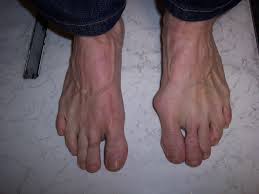
 "Until I met Jessica, she was in denial about her feet bubions, but after meeting her and checking out her bare feet, it was one of the first things I mentioned. Note to men : this is a really poor icebreaker!
"But I was forgiven, and I was right. Her shoes, even her most comfortable pair, were pushing her toes inward, particularly squeezing her biggest and smallest toes."
"Until I met Jessica, she was in denial about her feet bubions, but after meeting her and checking out her bare feet, it was one of the first things I mentioned. Note to men : this is a really poor icebreaker!
"But I was forgiven, and I was right. Her shoes, even her most comfortable pair, were pushing her toes inward, particularly squeezing her biggest and smallest toes."

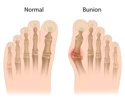 This is not the natural shape of the foot, and bunions are not hereditary. Instead, it's our feet directly adapting to the shape of our environment.


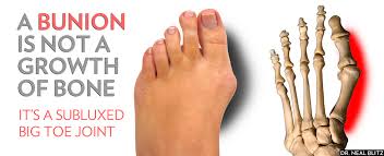
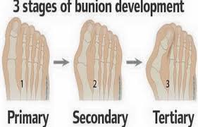 But why the bunion? The bony callus on the side of your foot is a protective mechanism, trying to get the shoe off your toes to give the toes more room to breathe and move properly.Over time that bunion can become quite painful if you don't heed its warning and give your feet the room they need.
Bunions, however are reversible! Once you begin taking your feet out of their medieval torture devices - your shoes, your feet begin to return to their natural shape, without bunions. Walking barefoot can be corrective for bunions in helping wake up the foot.
But why the bunion? The bony callus on the side of your foot is a protective mechanism, trying to get the shoe off your toes to give the toes more room to breathe and move properly.Over time that bunion can become quite painful if you don't heed its warning and give your feet the room they need.
Bunions, however are reversible! Once you begin taking your feet out of their medieval torture devices - your shoes, your feet begin to return to their natural shape, without bunions. Walking barefoot can be corrective for bunions in helping wake up the foot.
As Dr. Ray McClanahan, a leading podiatrist in the Northwest, put it this way, foot surgery, like that for bunions, typically isn't corrective surgery, but cosmetic surgery, done to help you feet fit into fashionable footwear. If you are willing to go barefoot, you can help correct your bunion naturally for free.

 The skeletal structure displaying various pathologies.
The function of all of the muscles in the lower limb originating below the knee is to move the foot. These muscles insert on any one or several of the 26 bones of the foot.
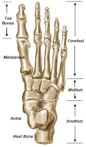
Bones of the Foot
The 26 bones of the foot, separated into the three main sections of the foot: Forefoot, Midfoot, Hindfoot, is summarized below:
 (From toes to heel) ⇧
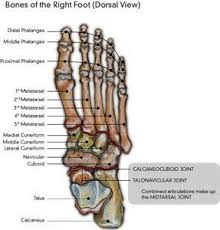
Bones of the Right Foot
Distal Phalanges
Middle Phalanges
Proximal Phalanges
(From toes to heel) ⇧
Bones of the Right Foot
Distal Phalanges
Middle Phalanges
Proximal Phalanges
1st Metatarsal
2nd Metatarsal
3rd Metatarsal
4th Metatarsal
5th Metatarsal
Medial Cuneiform
Middle Cuneiform
Lateral Cuneiform
Navicular
Cuboid
CALCANEOCUBOID JOINT
TALONAVICULAR JOINT
Combined articulations make up
the MIDTARSAL JOINT
Talus
Calcaneus

⇧Bones of Right Foot (Lateral View)
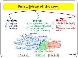
Joints of the Foot

The forefoot.
The forefoot consists of five metatarsals starting with the first to the fifth; and five toes, each of which consists of three bones (except for the big toe which consists of two). The bones of
each toe are the proximal phalanx, the middle phalanx, and the distal phalanx (except the big toe which has only proximal and distal). Between each of these bones is a joint which allows for the
movement necessary of each section of the foot.
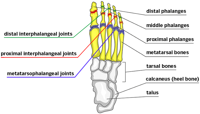
The joints of the forefoot are:
1. MTP joint - metatarsal phalangeal joint - between the metatarsal and the proximal phalanx of the adjacent toe.
2. PIP joint - proximal interphalangeal joint - between the proximal phalanx and the middle phalanx of each toe.
3. DIP joint - distal interphalangeal joint - between the middle phalanx and the distal phalanx of each toe.
4. The big (“great”) toe has only one joint between its two phalanges and therefore this joint is called the great (or “big” ) toe interphalangeal joint.
Metatarsal head is the end of the metatarsals, which articulate with the joints of the adjacent bones (generally used to describe the distal metatarsal head, the portion that articulates with the proximal
phalanx of the adjacent toe.)

The midfoot:
The Midfoot consists of five bones with numerous articular surfaces (surfaces which articulate by
way of joints with other bones).
1. navicular
2. cuboid
3. three cuneiform bones: medial, middle and lateral
Distally, the fourth and fifth metatarsals articulate with the cuboid bone. The first, second and third metatarsals articulate with each of their respective cuneiform bones. Each of these has an
individual joint capsule but all are wrapped in one big capsule as well to form the tarso-metatarsal joint (the “Lis Franc joint”).

 Proximally, the talonavicular and calcaneocuboid joints, together form the combined articulations of the midtarsal joint (of “Chopart”).
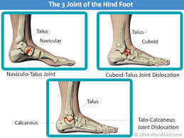
 The Hindfoot
The tibia articulates with the dome of the talus and thereby transmits the forces of the leg to the ankle. This is commonly called the “Tibialtalar joint” or simply the “Ankle joint”. In turn, the
talus articulates with the calcaneus, the main weight-bearing (and the largest) bone of the foot by way of the subtalar joint.
The Hindfoot
The tibia articulates with the dome of the talus and thereby transmits the forces of the leg to the ankle. This is commonly called the “Tibialtalar joint” or simply the “Ankle joint”. In turn, the
talus articulates with the calcaneus, the main weight-bearing (and the largest) bone of the foot by way of the subtalar joint.
 The subtalar joint, known as the “agility joint”, is a key joint in the ankle. It has three surfaces of articulation with three separate facet joints. A great deal of the movement in the ankle happens in this joint - the rest of the movement happens at the tibialtalar joint.
The plantar fascia is an important stabilizer in the foot where a great deal of foot pathology begins. The plantar fascia originates from the plantar surface of the calcaneus and attaches to the plantar
surfaces of the five metatarsal heads and proximal phalanges of the toes. The plantar fascia acts as a major stabilizer of the foot (see diagram). It helps maintain the arch of the foot and is an antipronator. In its function of maintaining the congruity of the relationship between the calcaneus and the metatarsal heads, it resists the torsion movement of the forefoot in relation to the hindfoot during pronation. Most of the eversion of pronation occurs in the mid and forefoot while the calcaneus remains stable in the hindfoot.
http://www.wefixfeet.ca/sites/default/files/anatomyofthefoot.pdf
