Tuesday, August 6, 2019

Water Balance

Water Balance


Water Balance

Chapter 26. Fluid, Electrolyte, and Acid-Base Balance

182 26.2 Water Balance

Learning Objectives

By the end of this section, you will be able to:
  • Explain how water levels in the body influence the thirst cycle
  • Identify the main route by which water leaves the body
  • Describe the role of ADH and its effect on body water levels
  • Define dehydration and identify common causes of dehydration
On a typical day, the average adult will take in about 2500 mL (almost 3 quarts) of aqueous fluids. Although most of the intake comes through the digestive tract, about 230 mL (8 ounces) per day is generated metabolically, in the last steps of aerobic respiration. Additionally, each day about the same volume (2500 mL) of water leaves the body by different routes; most of this lost water is removed as urine. The kidneys also can adjust blood volume though mechanisms that draw water out of the filtrate and urine. The kidneys can regulate water levels in the body; they conserve water if you are dehydrated, and they can make urine more dilute to expel excess water if necessary. Water is lost through the skin through evaporation from the skin surface without overt sweating and from air expelled from the lungs. This type of water loss is called insensible water loss because a person is usually unaware of it.

Regulation of Water Intake

Osmolality is the ratio of solutes in a solution to a volume of solvent in a solution. Plasma osmolality is thus the ratio of solutes to water in blood plasma. A person’s plasma osmolality value reflects his or her state of hydration. A healthy body maintains plasma osmolality within a narrow range, by employing several mechanisms that regulate both water intake and output.
Drinking water is considered voluntary. So how is water intake regulated by the body? Consider someone who is experiencing dehydration, a net loss of water that results in insufficient water in blood and other tissues. The water that leaves the body, as exhaled air, sweat, or urine, is ultimately extracted from blood plasma. As the blood becomes more concentrated, the thirst response—a sequence of physiological processes—is triggered (Figure 1). Osmoreceptors are sensory receptors in the thirst center in the hypothalamus that monitor the concentration of solutes (osmolality) of the blood. If blood osmolality increases above its ideal value, the hypothalamus transmits signals that result in a conscious awareness of thirst. The person should (and normally does) respond by drinking water. The hypothalamus of a dehydrated person also releases antidiuretic hormone (ADH) through the posterior pituitary gland. ADH signals the kidneys to recover water from urine, effectively diluting the blood plasma. To conserve water, the hypothalamus of a dehydrated person also sends signals via the sympathetic nervous system to the salivary glands in the mouth. The signals result in a decrease in watery, serous output (and an increase in stickier, thicker mucus output). These changes in secretions result in a “dry mouth” and the sensation of thirst.
This figure is a top-to bottom flowchart describing the thirst response. The topmost box of the chart states that there is insufficient water in the body, which has two effects. The left branch of the chart leads to decreased blood volume, which leads to decreased blood pressure. This triggers an increase in angiotensin two. Angiotensin two stimulates the thirst center in the hypothalamus. On the right branch, insufficient water in the body leads to increased blood osmolality, which causes dry mouth. Increased blood osmolality and dry mouth is sensed by osmoreceptors in the hypothalamus. This stimulates the thirst center in the hypothalamus to increase thirst, giving a person the urge to drink. Drinking decreases blood osmolality back to homeostatic levels.
Figure 1. A Flowchart Showing the Thirst Response. The thirst response begins when osmoreceptors detect a decrease in water levels in the blood.
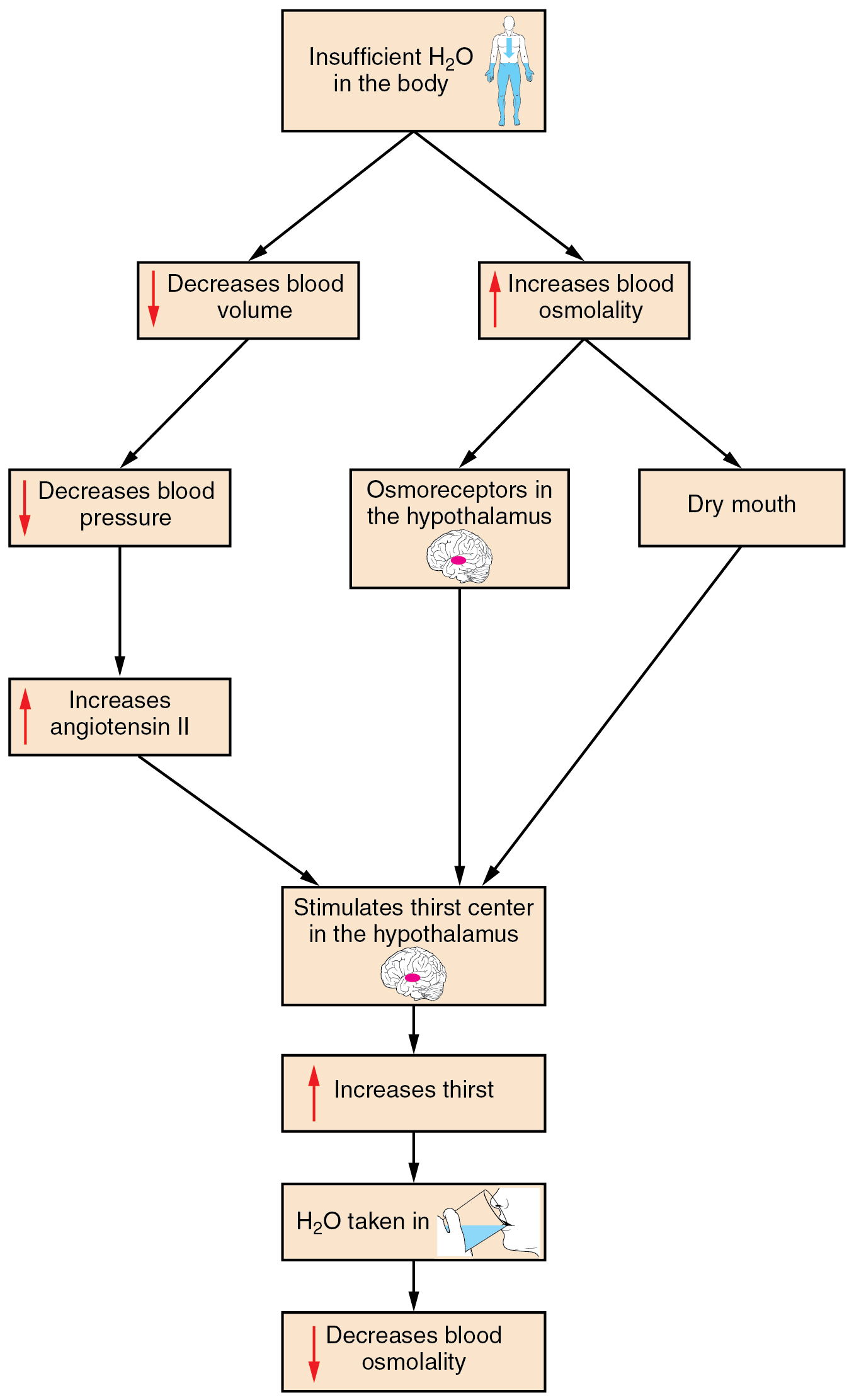
Decreased blood volume resulting from water loss has two additional effects. First, baroreceptors, blood-pressure receptors in the arch of the aorta and the carotid arteries in the neck, detect a decrease in blood pressure that results from decreased blood volume. The heart is ultimately signaled to increase its rate and/or strength of contractions to compensate for the lowered blood pressure.
Second, the kidneys have a renin-angiotensin hormonal system that increases the production of the active form of the hormone angiotensin II, which helps stimulate thirst, but also stimulates the release of the hormone aldosterone from the adrenal glands. Aldosterone increases the reabsorption of sodium in the distal tubules of the nephrons in the kidneys, and water follows this reabsorbed sodium back into the blood.
If adequate fluids are not consumed, dehydration results and a person’s body contains too little water to function correctly. A person who repeatedly vomits or who has diarrhea may become dehydrated, and infants, because their body mass is so low, can become dangerously dehydrated very quickly. Endurance athletes such as distance runners often become dehydrated during long races. Dehydration can be a medical emergency, and a dehydrated person may lose consciousness, become comatose, or die, if his or her body is not rehydrated quickly.

Regulation of Water Output

Water loss from the body occurs predominantly through the renal system. A person produces an average of 1.5 liters (1.6 quarts) of urine per day. Although the volume of urine varies in response to hydration levels, there is a minimum volume of urine production required for proper bodily functions. The kidney excretes 100 to 1200 milliosmoles of solutes per day to rid the body of a variety of excess salts and other water-soluble chemical wastes, most notably creatinine, urea, and uric acid. Failure to produce the minimum volume of urine means that metabolic wastes cannot be effectively removed from the body, a situation that can impair organ function. The minimum level of urine production necessary to maintain normal function is about 0.47 liters (0.5 quarts) per day.
The kidneys also must make adjustments in the event of ingestion of too much fluid. Diuresis, which is the production of urine in excess of normal levels, begins about 30 minutes after drinking a large quantity of fluid. Diuresis reaches a peak after about 1 hour, and normal urine production is reestablished after about 3 hours.

Role of ADH

Antidiuretic hormone (ADH), also known as vasopressin, controls the amount of water reabsorbed from the collecting ducts and tubules in the kidney. This hormone is produced in the hypothalamus and is delivered to the posterior pituitary for storage and release (Figure 2). When the osmoreceptors in the hypothalamus detect an increase in the concentration of blood plasma, the hypothalamus signals the release of ADH from the posterior pituitary into the blood.
This set of diagrams shows the effects of ADH on various structures within the body. In the brain, ADH affects the cerebrum by influencing social behavior in some mammals. ADH is also produced in the brain by the hypothalamus and released in the posterior pituitary. ADH also constricts arterioles in the body, which are the small arteries that enter into capillary beds. Finally, a kidney is shown because ADH increases the reabsorption of water in the kidneys.
Figure 2. Antidiuretic Hormone (ADH). ADH is produced in the hypothalamus and released by the posterior pituitary gland. It causes the kidneys to retain water, constricts arterioles in the peripheral circulation, and affects some social behaviors in mammals.
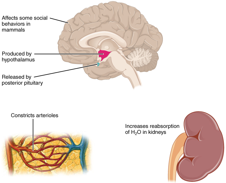
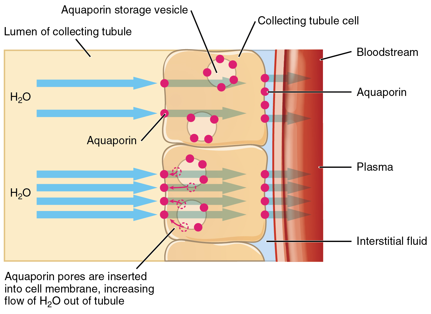
ADH has two major effects. It constricts the arterioles in the peripheral circulation, which reduces the flow of blood to the extremities and thereby increases the blood supply to the core of the body. ADH also causes the epithelial cells that line the renal collecting tubules to move water channel proteins, called aquaporins, from the interior of the cells to the apical surface, where these proteins are inserted into the cell membrane (Figure 3). The result is an increase in the water permeability of these cells and, thus, a large increase in water passage from the urine through the walls of the collecting tubules, leading to more reabsorption of water into the bloodstream. When the blood plasma becomes less concentrated and the level of ADH decreases, aquaporins are removed from collecting tubule cell membranes, and the passage of water out of urine and into the blood decreases.
This diagram depicts a cross section of the right wall of a kidney collecting tubule. The wall is composed of three block-shaped cells arranged vertically one on top of each other. The lumen of the collecting tubule is to the left of the three cells. Yellow-colored urine is flowing through the lumen. There is a small strip of blue interstitial fluid to the right of the three cells. To the right of the interstitial fluid is a cross section of a blood vessel. Arrows show that water in the urine is entering the left side of the wall cells through aquaporins. The water travels through the cells and then leaves the kidney tubule through additional aquaporins in the right side of the wall cells. The water travels through the interstitial space and enters into the blood in the blood vessel. The aquaporins in the wall cells are being released from aquaporin storage vesicles within their cytoplasm.
Figure 3. Aquaporins. The binding of ADH to receptors on the cells of the collecting tubule results in aquaporins being inserted into the plasma membrane, shown in the lower cell. This dramatically increases the flow of water out of the tubule and into the bloodstream.
A diuretic is a compound that increases urine output and therefore decreases water conservation by the body. Diuretics are used to treat hypertension, congestive heart failure, and fluid retention associated with menstruation. Alcohol acts as a diuretic by inhibiting the release of ADH. Additionally, caffeine, when consumed in high concentrations, acts as a diuretic.

Chapter Review

Homeostasis requires that water intake and output be balanced. Most water intake comes through the digestive tract via liquids and food, but roughly 10 percent of water available to the body is generated at the end of aerobic respiration during cellular metabolism. Urine produced by the kidneys accounts for the largest amount of water leaving the body. The kidneys can adjust the concentration of the urine to reflect the body’s water needs, conserving water if the body is dehydrated or making urine more dilute to expel excess water when necessary. ADH is a hormone that helps the body to retain water by increasing water reabsorption by the kidneys.

Review Questions

1. The largest amount of water comes into the body via ________.
  1. metabolism
  2. foods
  3. liquids
  4. humidified air
2. The largest amount of water leaves the body via ________.
  1. the GI tract
  2. the skin as sweat
  3. expiration
  4. urine
3. Insensible water loss is water lost via ________.
  1. skin evaporation and in air from the lungs
  2. urine
  3. excessive sweating
  4. vomiting or diarrhea
4. How soon after drinking a large glass of water will a person start increasing their urine output?
  1. 5 minutes
  2. 30 minutes
  3. 1 hour
  4. 3 hours

Critical Thinking Questions

1. Describe the effect of ADH on renal collecting tubules.
2. Why is it important for the amount of water intake to equal the amount of water output?

Glossary

antidiuretic hormone (ADH)
also known as vasopressin, a hormone that increases the volume of water reabsorbed from the collecting tubules of the kidney
dehydration
state of containing insufficient water in blood and other tissues
diuresis
excess production of urine
plasma osmolality
ratio of solutes to a volume of solvent in the plasma; plasma osmolality reflects a person’s state of hydration

Solutions

Answers for Review Questions
  1. C
  2. D
  3. A
  4. B
Answers for Critical Thinking Questions
  1. ADH constricts the arterioles in the peripheral circulation, limiting blood to the extremities and increasing the blood supply to the core of the body. ADH also causes the epithelial cells lining the renal collecting tubules to move water channel proteins called aquaporins from the sides of the cells to the apical surface. This greatly increases the passage of water from the renal filtrate through the wall of the collecting tubule as well as the reabsorption of water into the bloodstream.
  2. Any imbalance of water entering or leaving the body will create an osmotic imbalance that will adversely affect cell and tissue function.

Chapter 26. Fluid, Electrolyte, and Acid-Base Balance

Chapter 26. Fluid, Electrolyte, and Acid-Base Balance

180 Introduction
This is a photo of Venus Williams, the famous tennis player, executing a hard tennis swing.
Figure 1. Venus Williams Perspiring on the Tennis Court. The body has critically important mechanisms for balancing the intake and output of bodily fluids. An athlete must continuously replace the water and electrolytes lost in sweat. (credit: “Edwin Martinez1”/Wikimedia Commons)
Chapter Objectives
After studying this chapter, you will be able to:

Identify the body’s main fluid compartments
Define plasma osmolality and identify two ways in which plasma osmolality is maintained
Identify the six ions most important to the function of the body
Define buffer and discuss the role of buffers in the body
Explain why bicarbonate must be conserved rather than reabsorbed in the kidney
Identify the normal range of blood pH and name the conditions where one has a blood pH that is either too high or too low

Homeostasis, or the maintenance of constant conditions in the body, is a fundamental property of all living things. In the human body, the substances that participate in chemical reactions must remain within narrows ranges of concentration. Too much or too little of a single substance can disrupt your bodily functions. Because metabolism relies on reactions that are all interconnected, any disruption might affect multiple organs or even organ systems. Water is the most ubiquitous substance in the chemical reactions of life. The interactions of various aqueous solutions—solutions in which water is the solvent—are continuously monitored and adjusted by a large suite of interconnected feedback systems in your body. Understanding the ways in which the body maintains these critical balances is key to understanding good health.

180 Introduction

This is a photo of Venus Williams, the famous tennis player, executing a hard tennis swing.
Figure 1. Venus Williams Perspiring on the Tennis Court. The body has critically important mechanisms for balancing the intake and output of bodily fluids. An athlete must continuously replace the water and electrolytes lost in sweat. (credit: “Edwin Martinez1”/Wikimedia Commons)

Chapter Objectives

After studying this chapter, you will be able to:
  • Identify the body’s main fluid compartments
  • Define plasma osmolality and identify two ways in which plasma osmolality is maintained
  • Identify the six ions most important to the function of the body
  • Define buffer and discuss the role of buffers in the body
  • Explain why bicarbonate must be conserved rather than reabsorbed in the kidney
  • Identify the normal range of blood pH and name the conditions where one has a blood pH that is either too high or too low
Homeostasis, or the maintenance of constant conditions in the body, is a fundamental property of all living things. In the human body, the substances that participate in chemical reactions must remain within narrows ranges of concentration. Too much or too little of a single substance can disrupt your bodily functions. Because metabolism relies on reactions that are all interconnected, any disruption might affect multiple organs or even organ systems. Water is the most ubiquitous substance in the chemical reactions of life. The interactions of various aqueous solutions—solutions in which water is the solvent—are continuously monitored and adjusted by a large suite of interconnected feedback systems in your body. Understanding the ways in which the body maintains these critical balances is key to understanding good health.


Body Fluids and Fluid Compartments

 Body Fluids and Fluid Compartments


 Body Fluids and Fluid Compartments

Learning Objectives

By the end of this section, you will be able to:

Explain the importance of water in the body.
Contrast the composition of the intracellular fluid with that of the extracellular fluid.
Explain the importance of protein channels in the movement of solutes.
Identify the causes and symptoms of edema.

The chemical reactions of life take place in aqueous solutions. The dissolved substances in a solution are called solutes. In the human body, solutes vary in different parts of the body, but may include proteins—including those that transport lipids, carbohydrates, and, very importantly, electrolytes. Often in medicine, a mineral dissociated from a salt that carries an electrical charge (an ion) is called and electrolyte. For instance, sodium ions (Na+) and chloride ions (Cl–) are often referred to as electrolytes.

In the body, water moves through semi-permeable membranes of cells and from one compartment of the body to another by a process called osmosis. Osmosis is basically the diffusion of water from regions of higher concentration to regions of lower concentration, along an osmotic gradient across a semi-permeable membrane. As a result, water will move into and out of cells and tissues, depending on the relative concentrations of the water and solutes found there. An appropriate balance of solutes inside and outside of cells must be maintained to ensure normal function.

Body Water Content
Human beings are mostly water, ranging from about 75 percent of body mass in infants to about 50–60 percent in adult men and women, to as low as 45 percent in old age. The percent of body water changes with development, because the proportions of the body given over to each organ and to muscles, fat, bone, and other tissues change from infancy to adulthood (Figure 1). Your brain and kidneys have the highest proportions of water, which composes 80–85 percent of their masses. In contrast, teeth have the lowest proportion of water, at 8–10 percent.

This illustration shows a silhouette of a human body with various organs highlighted. The percent of water contained in each organ is given. The brain typically contains 80% to 85% water, teeth contain 8% to 10% water, a single lung contains 75% to 80% water, the heart contains 75% to 80% water, the bones contain 20% to 25% water, the liver contains 70% to 75% water, the kidneys contain 80% to 85% water, the skin contains 70% to 75% water and the muscles also contain 70% to 75% water.

Figure 1. Water Content of the Body’s Organs and Tissues. Water content varies in different body organs and tissues, from as little as 8 percent in the teeth to as much as 85 percent in the brain.

Fluid Compartments

Body fluids can be discussed in terms of their specific fluid compartment, a location that is largely separate from another compartment by some form of a physical barrier. The intracellular fluid (ICF) compartment is the system that includes all fluid enclosed in cells by their plasma membranes. Extracellular fluid (ECF) surrounds all cells in the body. Extracellular fluid has two primary constituents: the fluid component of the blood (called plasma) and the interstitial fluid (IF) that surrounds all cells not in the blood (Figure 2).

This diagram shows a small blood vessel surrounded by several body cells. The fluid between the body cells is the interstitial fluid (IF), which is a type of extracellular fluid (ECF). The fluid in the blood vessel is also an example of extracellular fluid. The fluid in the cytoplasm of each body cell is intracellular fluid, or ICF.

Figure 2. Fluid Compartments in the Human Body. The intracellular fluid (ICF) is the fluid within cells. The interstitial fluid (IF) is part of the extracellular fluid (ECF) between the cells. Blood plasma is the second part of the ECF. Materials travel between cells and the plasma in capillaries through the IF.

Intracellular Fluid

The ICF lies within cells and is the principal component of the cytosol/cytoplasm. The ICF makes up about 60 percent of the total water in the human body, and in an average-size adult male, the ICF accounts for about 25 liters (seven gallons) of fluid (Figure 3). This fluid volume tends to be very stable, because the amount of water in living cells is closely regulated. If the amount of water inside a cell falls to a value that is too low, the cytosol becomes too concentrated with solutes to carry on normal cellular activities; if too much water enters a cell, the cell may burst and be destroyed.

This pie chart shows that about 55% of water in the human body is intracellular fluid. About 30% of the water in the human body is interstitial fluid. Most of the remaining 15% of water is plasma, along with a small percentage labeled “other fluid”.

Figure 3. A Pie Graph Showing the Proportion of Total Body Fluid in Each of the Body’s Fluid Compartments. Most of the water in the body is intracellular fluid. The second largest volume is the interstitial fluid, which surrounds cells that are not blood cells.

Extracellular Fluid
The ECF accounts for the other one-third of the body’s water content. Approximately 20 percent of the ECF is found in plasma. Plasma travels through the body in blood vessels and transports a range of materials, including blood cells, proteins (including clotting factors and antibodies), electrolytes, nutrients, gases, and wastes. Gases, nutrients, and waste materials travel between capillaries and cells through the IF. Cells are separated from the IF by a selectively permeable cell membrane that helps regulate the passage of materials between the IF and the interior of the cell.

The body has other water-based ECF. These include the cerebrospinal fluid that bathes the brain and spinal cord, lymph, the synovial fluid in joints, the pleural fluid in the pleural cavities, the pericardial fluid in the cardiac sac, the peritoneal fluid in the peritoneal cavity, and the aqueous humor of the eye. Because these fluids are outside of cells, these fluids are also considered components of the ECF compartment.

Composition of Body Fluids

The compositions of the two components of the ECF—plasma and IF—are more similar to each other than either is to the ICF (Figure 4). Blood plasma has high concentrations of sodium, chloride, bicarbonate, and protein. The IF has high concentrations of sodium, chloride, and bicarbonate, but a relatively lower concentration of protein. In contrast, the ICF has elevated amounts of potassium, phosphate, magnesium, and protein. Overall, the ICF contains high concentrations of potassium and phosphate (HPO42−HPO42−), whereas both plasma and the ECF contain high concentrations of sodium and chloride.

This bar graph shows the concentration of several ions and proteins in intracellular fluid, interstitial fluid and plasma. The ions and proteins are categories on the X axis . The Y axis shows concentration, in milliequivalents per liter, ranging from zero to 160. Three different colored bars are shown above each compound on the X axis. One bar represents intracellular fluid (ICF), a second bar represents interstitial fluid (IF, which is part of ECF) and the third bar represents plasma (ECF). Intracellular fluid contains high concentrations of K plus and HPO four two minus. It has lower concentrations of MG two plus and protein, and negligible amounts of the other compounds. Interstitial fluid contains high concentrations of NA plus and CL minus, along with a smaller amount of HCO 3 minus, and negligible amounts of the other compounds. Plasma contains large concentrations of NA plus and CL minus, with smaller concentrations of HCO 3 minus and protein, and negligible amounts of the other compounds.

Figure 4. The Concentrations of Different Elements in Key Bodily Fluids. The graph shows the composition of the ICF, IF, and plasma. The compositions of plasma and IF are similar to one another but are quite different from the composition of the ICF.

QR Codes representing a URL

Watch this video (click here) to learn more about body fluids, fluid compartments, and electrolytes.
Watch this video to learn more about body fluids, fluid compartments, and electrolytes. When blood volume decreases due to sweating, from what source is water taken in by the blood?

Most body fluids are neutral in charge. Thus, cations, or positively charged ions, and anions, or negatively charged ions, are balanced in fluids. As seen in the previous graph, sodium (Na+) ions and chloride (Cl–) ions are concentrated in the ECF of the body, whereas potassium (K+) ions are concentrated inside cells. Although sodium and potassium can “leak” through “pores” into and out of cells, respectively, the high levels of potassium and low levels of sodium in the ICF are maintained by sodium-potassium pumps in the cell membranes. These pumps use the energy supplied by ATP to pump sodium out of the cell and potassium into the cell (Figure 5).

This diagram shows a sodium potassium pump embedded in the cell membrane. In the first step, the pump is opened to the cytosol and closed to the extracellular fluid. First, three sodium ions move into the pump from the cytosol. An ATP molecule binds to the cytosol side of the pump, causing the pump to change shape and open to the extracellular fluid. The pump is now closed to the cytosol. The sodium ions are then released into the extracellular fluid, after which two potassium ions enter the pump. Also at this point, the used ADP detaches from the cytosol side of the pump, leaving a single phosphate attached. The pump then changes shape again so that it closes to the extracellular fluid and again opens to the cytosol. This releases the two potassium ions into the cytosol. The single phosphate also detaches from the pump at this point so that the cycle can start anew. Two bars along the right hand side of the figure indicate that sodium normally diffuses into the cell down its concentration gradient while potassium usually diffuses out of the cell down its concentration gradient. Therefore, the sodium potassium pump is working against these natural concentration gradients.

Figure 5. The Sodium-Potassium Pump. The sodium-potassium pump is powered by ATP to transfer sodium out of the cytoplasm and into the ECF. The pump also transfers potassium out of the ECF and into the cytoplasm. (credit: modification of work by Mariana Ruiz Villarreal)

Fluid Movement between Compartments

Hydrostatic pressure, the force exerted by a fluid against a wall, causes movement of fluid between compartments. The hydrostatic pressure of blood is the pressure exerted by blood against the walls of the blood vessels by the pumping action of the heart. In capillaries, hydrostatic pressure (also known as capillary blood pressure) is higher than the opposing “colloid osmotic pressure” in blood—a “constant” pressure primarily produced by circulating albumin—at the arteriolar end of the capillary (Figure 6). This pressure forces plasma and nutrients out of the capillaries and into surrounding tissues. Fluid and the cellular wastes in the tissues enter the capillaries at the venule end, where the hydrostatic pressure is less than the osmotic pressure in the vessel. Filtration pressure squeezes fluid from the plasma in the blood to the IF surrounding the tissue cells. The surplus fluid in the interstitial space that is not returned directly back to the capillaries is drained from tissues by the lymphatic system, and then re-enters the vascular system at the subclavian veins.


Figure 6. Capillary Exchange. Net filtration occurs near the arterial end of the capillary since capillary hydrostatic pressure (CHP) is greater than blood colloidal osmotic pressure (BCOP). There is no net movement of fluid near the midpoint of the capillary since CHP = BCOP. Net reabsorption occurs near the venous end of the capillary since BCOP is greater than CHP.

QR Code representing a URL

Watch this video to see an explanation of the dynamics of fluid in the body’s compartments.
Watch this video to see an explanation of the dynamics of fluid in the body’s compartments. What happens in the tissue when capillary blood pressure is less than osmotic pressure?

Hydrostatic pressure is especially important in governing the movement of water in the nephrons of the kidneys to ensure proper filtering of the blood to form urine. As hydrostatic pressure in the kidneys increases, the amount of water leaving the capillaries also increases, and more urine filtrate is formed. If hydrostatic pressure in the kidneys drops too low, as can happen in dehydration, the functions of the kidneys will be impaired, and less nitrogenous wastes will be removed from the bloodstream. Extreme dehydration can result in kidney failure.

Fluid also moves between compartments along an osmotic gradient. Recall that an osmotic gradient is produced by the difference in concentration of all solutes on either side of a semi-permeable membrane. The magnitude of the osmotic gradient is proportional to the difference in the concentration of solutes on one side of the cell membrane to that on the other side. Water will move by osmosis from the side where its concentration is high (and the concentration of solute is low) to the side of the membrane where its concentration is low (and the concentration of solute is high). In the body, water moves by osmosis from plasma to the IF (and the reverse) and from the IF to the ICF (and the reverse). In the body, water moves constantly into and out of fluid compartments as conditions change in different parts of the body.

For example, if you are sweating, you will lose water through your skin. Sweating depletes your tissues of water and increases the solute concentration in those tissues. As this happens, water diffuses from your blood into sweat glands and surrounding skin tissues that have become dehydrated because of the osmotic gradient. Additionally, as water leaves the blood, it is replaced by the water in other tissues throughout your body that are not dehydrated. If this continues, dehydration spreads throughout the body. When a dehydrated person drinks water and rehydrates, the water is redistributed by the same gradient, but in the opposite direction, replenishing water in all of the tissues.

Solute Movement between Compartments

The movement of some solutes between compartments is active, which consumes energy and is an active transport process, whereas the movement of other solutes is passive, which does not require energy. Active transport allows cells to move a specific substance against its concentration gradient through a membrane protein, requiring energy in the form of ATP. For example, the sodium-potassium pump employs active transport to pump sodium out of cells and potassium into cells, with both substances moving against their concentration gradients.

Passive transport of a molecule or ion depends on its ability to pass through the membrane, as well as the existence of a concentration gradient that allows the molecules to diffuse from an area of higher concentration to an area of lower concentration. Some molecules, like gases, lipids, and water itself (which also utilizes water channels in the membrane called aquaporins), slip fairly easily through the cell membrane; others, including polar molecules like glucose, amino acids, and ions do not. Some of these molecules enter and leave cells using facilitated transport, whereby the molecules move down a concentration gradient through specific protein channels in the membrane. This process does not require energy. For example, glucose is transferred into cells by glucose transporters that use facilitated transport (Figure 7).

This diagram shows a carrier protein embedded in the plasma membrane between the cytoplasm and the extracellular fluid. There are several glucose molecules in the extracellular fluid. In the first step, the carrier protein is open to the extracellular fluid and closed to the cytosol. One of the glucose molecules travels from the extracellular fluid into the carrier protein. The protein then changes shape, closing at both ends. This pushes the glucose down into the carrier protein, closer to the cytosol end. The protein then opens on the cytosol side and closes on the extracellular fluid side, allowing the glucose to enter the cytosol.
Figure 7. Facilitated Diffusion. Glucose molecules use facilitated diffusion to move down a concentration gradient through the carrier protein channels in the membrane. (credit: modification of work by Mariana Ruiz Villarreal)

Disorders of the…

Fluid Balance: Edema

Edema is the accumulation of excess water in the tissues. It is most common in the soft tissues of the extremities. The physiological causes of edema include water leakage from blood capillaries. Edema is almost always caused by an underlying medical condition, by the use of certain therapeutic drugs, by pregnancy, by localized injury, or by an allergic reaction. In the limbs, the symptoms of edema include swelling of the subcutaneous tissues, an increase in the normal size of the limb, and stretched, tight skin. One quick way to check for subcutaneous edema localized in a limb is to press a finger into the suspected area. Edema is likely if the depression persists for several seconds after the finger is removed (which is called “pitting”).

Pulmonary edema is excess fluid in the air sacs of the lungs, a common symptom of heart and/or kidney failure. People with pulmonary edema likely will experience difficulty breathing, and they may experience chest pain. Pulmonary edema can be life threatening, because it compromises gas exchange in the lungs, and anyone having symptoms should immediately seek medical care.

In pulmonary edema resulting from heart failure, excessive leakage of water occurs because fluids get “backed up” in the pulmonary capillaries of the lungs, when the left ventricle of the heart is unable to pump sufficient blood into the systemic circulation. Because the left side of the heart is unable to pump out its normal volume of blood, the blood in the pulmonary circulation gets “backed up,” starting with the left atrium, then into the pulmonary veins, and then into pulmonary capillaries. The resulting increased hydrostatic pressure within pulmonary capillaries, as blood is still coming in from the pulmonary arteries, causes fluid to be pushed out of them and into lung tissues.

Other causes of edema include damage to blood vessels and/or lymphatic vessels, or a decrease in osmotic pressure in chronic and severe liver disease, where the liver is unable to manufacture plasma proteins (Figure 8). A decrease in the normal levels of plasma proteins results in a decrease of colloid osmotic pressure (which counterbalances the hydrostatic pressure) in the capillaries. This process causes loss of water from the blood to the surrounding tissues, resulting in edema.

This photo shows the dorsal surfaces of a person’s right and left hands. The left hand is normal, with the several blood vessels visible under the skin. However, the top of the right hand is swollen and no blood vessels are visible.

Figure 8. Edema. An allergic reaction can cause capillaries in the hand to leak excess fluid that accumulates in the tissues. (credit: Jane Whitney)

Mild, transient edema of the feet and legs may be caused by sitting or standing in the same position for long periods of time, as in the work of a toll collector or a supermarket cashier. This is because deep veins in the lower limbs rely on skeletal muscle contractions to push on the veins and thus “pump” blood back to the heart. Otherwise, the venous blood pools in the lower limbs and can leak into surrounding tissues.

Medications that can result in edema include vasodilators, calcium channel blockers used to treat hypertension, non-steroidal anti-inflammatory drugs, estrogen therapies, and some diabetes medications. Underlying medical conditions that can contribute to edema include congestive heart failure, kidney damage and kidney disease, disorders that affect the veins of the legs, and cirrhosis and other liver disorders.

Therapy for edema usually focuses on elimination of the cause. Activities that can reduce the effects of the condition include appropriate exercises to keep the blood and lymph flowing through the affected areas. Other therapies include elevation of the affected part to assist drainage, massage and compression of the areas to move the fluid out of the tissues, and decreased salt intake to decrease sodium and water retention.

Chapter Review

Your body is mostly water. Body fluids are aqueous solutions with differing concentrations of materials, called solutes. An appropriate balance of water and solute concentrations must be maintained to ensure cellular functions. If the cytosol becomes too concentrated due to water loss, cell functions deteriorate. If the cytosol becomes too dilute due to water intake by cells, cell membranes can be damaged, and the cell can burst. Hydrostatic pressure is the force exerted by a fluid against a wall and causes movement of fluid between compartments. Fluid can also move between compartments along an osmotic gradient. Active transport processes require ATP to move some solutes against their concentration gradients between compartments. Passive transport of a molecule or ion depends on its ability to pass easily through the membrane, as well as the existence of a high to low concentration gradient.

Interactive Link Questions
Watch this video to learn more about body fluids, fluid compartments, and electrolytes. When blood volume decreases due to sweating, from what source is water taken in by the blood?

The interstitial fluid (IF).

Watch this video to see an explanation of the dynamics of fluid in the body’s compartments. What happens in tissues when capillary blood pressure is less than osmotic pressure?

Fluid enters the capillaries from interstitial spaces.

Review Questions
1. Solute contributes to the movement of water between cells and the surrounding medium by ________.

A. osmotic pressure
B. hydrostatic pressure
C. Brownian movement
D. random motion

2. A cation has a(n) ________ charge.

A. neutral
B. positive
C. alternating
D. negative

3. Interstitial fluid (IF) is ________.

A. the fluid in the cytosol of the cells
B. the fluid component of blood
C. the fluid that bathes all of the body’s cells except for blood cells
D. the intracellular fluids found between membranes

Critical Thinking Questions

1. Plasma contains more sodium than chloride. How can this be if individual ions of sodium and chloride exactly balance each other out, and plasma is electrically neutral?

2. How is fluid moved from compartment to compartment?

Glossary

extracellular fluid (ECF)
fluid exterior to cells; includes the interstitial fluid, blood plasma, and fluids found in other reservoirs in the body

fluid compartment
fluid inside all cells of the body constitutes a compartment system that is largely segregated from other systems

hydrostatic pressure
pressure exerted by a fluid against a wall, caused by its own weight or pumping force

interstitial fluid (IF)
fluid in the small spaces between cells not contained within blood vessels

intracellular fluid (ICF)
fluid in the cytosol of cells


Solutions
Answers for Review Questions

1. A
2. B
3. C


Answers for Critical Thinking Questions

1. There are additional negatively charged molecules in plasma besides chloride. The additional sodium balances the total negative charges.


2. Fluid is moved by a combination of osmotic and hydrostatic pressures. The osmotic pressure results from differences in solute concentrations across cell membranes. Hydrostatic pressure results from the pressure of blood as it enters a capillary system, forcing some fluid out of the vessel into the surrounding tissues.


Learning Objectives

By the end of this section, you will be able to:
  • Explain the importance of water in the body
  • Contrast the composition of the intracellular fluid with that of the extracellular fluid
  • Explain the importance of protein channels in the movement of solutes
  • Identify the causes and symptoms of edema
The chemical reactions of life take place in aqueous solutions. The dissolved substances in a solution are called solutes. In the human body, solutes vary in different parts of the body, but may include proteins—including those that transport lipids, carbohydrates, and, very importantly, electrolytes. Often in medicine, a mineral dissociated from a salt that carries an electrical charge (an ion) is called and electrolyte. For instance, sodium ions (Na+) and chloride ions (Cl) are often referred to as electrolytes.
In the body, water moves through semi-permeable membranes of cells and from one compartment of the body to another by a process called osmosis. Osmosis is basically the diffusion of water from regions of higher concentration to regions of lower concentration, along an osmotic gradient across a semi-permeable membrane. As a result, water will move into and out of cells and tissues, depending on the relative concentrations of the water and solutes found there. An appropriate balance of solutes inside and outside of cells must be maintained to ensure normal function.

Body Water Content

Human beings are mostly water, ranging from about 75 percent of body mass in infants to about 50–60 percent in adult men and women, to as low as 45 percent in old age. The percent of body water changes with development, because the proportions of the body given over to each organ and to muscles, fat, bone, and other tissues change from infancy to adulthood (Figure 1). Your brain and kidneys have the highest proportions of water, which composes 80–85 percent of their masses. In contrast, teeth have the lowest proportion of water, at 8–10 percent.
This illustration shows a silhouette of a human body with various organs highlighted. The percent of water contained in each organ is given. The brain typically contains 80% to 85% water, teeth contain 8% to 10% water, a single lung contains 75% to 80% water, the heart contains 75% to 80% water, the bones contain 20% to 25% water, the liver contains 70% to 75% water, the kidneys contain 80% to 85% water, the skin contains 70% to 75% water and the muscles also contain 70% to 75% water.
Figure 1. Water Content of the Body’s Organs and Tissues. Water content varies in different body organs and tissues, from as little as 8 percent in the teeth to as much as 85 percent in the brain.

Fluid Compartments

Body fluids can be discussed in terms of their specific fluid compartment, a location that is largely separate from another compartment by some form of a physical barrier. The intracellular fluid (ICF) compartment is the system that includes all fluid enclosed in cells by their plasma membranes. Extracellular fluid (ECF) surrounds all cells in the body. Extracellular fluid has two primary constituents: the fluid component of the blood (called plasma) and the interstitial fluid (IF) that surrounds all cells not in the blood (Figure 2).
This diagram shows a small blood vessel surrounded by several body cells. The fluid between the body cells is the interstitial fluid (IF), which is a type of extracellular fluid (ECF). The fluid in the blood vessel is also an example of extracellular fluid. The fluid in the cytoplasm of each body cell is intracellular fluid, or ICF.
Figure 2. Fluid Compartments in the Human Body. The intracellular fluid (ICF) is the fluid within cells. The interstitial fluid (IF) is part of the extracellular fluid (ECF) between the cells. Blood plasma is the second part of the ECF. Materials travel between cells and the plasma in capillaries through the IF.

Intracellular Fluid

The ICF lies within cells and is the principal component of the cytosol/cytoplasm. The ICF makes up about 60 percent of the total water in the human body, and in an average-size adult male, the ICF accounts for about 25 liters (seven gallons) of fluid (Figure 3). This fluid volume tends to be very stable, because the amount of water in living cells is closely regulated. If the amount of water inside a cell falls to a value that is too low, the cytosol becomes too concentrated with solutes to carry on normal cellular activities; if too much water enters a cell, the cell may burst and be destroyed.
This pie chart shows that about 55% of water in the human body is intracellular fluid. About 30% of the water in the human body is interstitial fluid. Most of the remaining 15% of water is plasma, along with a small percentage labeled “other fluid”.
Figure 3. A Pie Graph Showing the Proportion of Total Body Fluid in Each of the Body’s Fluid Compartments. Most of the water in the body is intracellular fluid. The second largest volume is the interstitial fluid, which surrounds cells that are not blood cells.

Extracellular Fluid

The ECF accounts for the other one-third of the body’s water content. Approximately 20 percent of the ECF is found in plasma. Plasma travels through the body in blood vessels and transports a range of materials, including blood cells, proteins (including clotting factors and antibodies), electrolytes, nutrients, gases, and wastes. Gases, nutrients, and waste materials travel between capillaries and cells through the IF. Cells are separated from the IF by a selectively permeable cell membrane that helps regulate the passage of materials between the IF and the interior of the cell.
The body has other water-based ECF. These include the cerebrospinal fluid that bathes the brain and spinal cord, lymph, the synovial fluid in joints, the pleural fluid in the pleural cavities, the pericardial fluid in the cardiac sac, the peritoneal fluid in the peritoneal cavity, and the aqueous humor of the eye. Because these fluids are outside of cells, these fluids are also considered components of the ECF compartment.

Composition of Body Fluids

The compositions of the two components of the ECF—plasma and IF—are more similar to each other than either is to the ICF (Figure 4). Blood plasma has high concentrations of sodium, chloride, bicarbonate, and protein. The IF has high concentrations of sodium, chloride, and bicarbonate, but a relatively lower concentration of protein. In contrast, the ICF has elevated amounts of potassium, phosphate, magnesium, and protein. Overall, the ICF contains high concentrations of potassium and phosphate (HPO42−HPO42−), whereas both plasma and the ECF contain high concentrations of sodium and chloride.
This bar graph shows the concentration of several ions and proteins in intracellular fluid, interstitial fluid and plasma. The ions and proteins are categories on the X axis . The Y axis shows concentration, in milliequivalents per liter, ranging from zero to 160. Three different colored bars are shown above each compound on the X axis. One bar represents intracellular fluid (ICF), a second bar represents interstitial fluid (IF, which is part of ECF) and the third bar represents plasma (ECF). Intracellular fluid contains high concentrations of K plus and HPO four two minus. It has lower concentrations of MG two plus and protein, and negligible amounts of the other compounds. Interstitial fluid contains high concentrations of NA plus and CL minus, along with a smaller amount of HCO 3 minus, and negligible amounts of the other compounds. Plasma contains large concentrations of NA plus and CL minus, with smaller concentrations of HCO 3 minus and protein, and negligible amounts of the other compounds.
Figure 4. The Concentrations of Different Elements in Key Bodily Fluids. The graph shows the composition of the ICF, IF, and plasma. The compositions of plasma and IF are similar to one another but are quite different from the composition of the ICF.

QR Codes representing a URL
Watch this video to learn more about body fluids, fluid compartments, and electrolytes.

Watch this video to learn more about body fluids, fluid compartments, and electrolytes. When blood volume decreases due to sweating, from what source is water taken in by the blood?
Most body fluids are neutral in charge. Thus, cations, or positively charged ions, and anions, or negatively charged ions, are balanced in fluids. As seen in the previous graph, sodium (Na+) ions and chloride (Cl) ions are concentrated in the ECF of the body, whereas potassium (K+) ions are concentrated inside cells. Although sodium and potassium can “leak” through “pores” into and out of cells, respectively, the high levels of potassium and low levels of sodium in the ICF are maintained by sodium-potassium pumps in the cell membranes. These pumps use the energy supplied by ATP to pump sodium out of the cell and potassium into the cell (Figure 5).
This diagram shows a sodium potassium pump embedded in the cell membrane. In the first step, the pump is opened to the cytosol and closed to the extracellular fluid. First, three sodium ions move into the pump from the cytosol. An ATP molecule binds to the cytosol side of the pump, causing the pump to change shape and open to the extracellular fluid. The pump is now closed to the cytosol. The sodium ions are then released into the extracellular fluid, after which two potassium ions enter the pump. Also at this point, the used ADP detaches from the cytosol side of the pump, leaving a single phosphate attached. The pump then changes shape again so that it closes to the extracellular fluid and again opens to the cytosol. This releases the two potassium ions into the cytosol. The single phosphate also detaches from the pump at this point so that the cycle can start anew. Two bars along the right hand side of the figure indicate that sodium normally diffuses into the cell down its concentration gradient while potassium usually diffuses out of the cell down its concentration gradient. Therefore, the sodium potassium pump is working against these natural concentration gradients.
Figure 5. The Sodium-Potassium Pump. The sodium-potassium pump is powered by ATP to transfer sodium out of the cytoplasm and into the ECF. The pump also transfers potassium out of the ECF and into the cytoplasm. (credit: modification of work by Mariana Ruiz Villarreal)

Fluid Movement between Compartments

Hydrostatic pressure, the force exerted by a fluid against a wall, causes movement of fluid between compartments. The hydrostatic pressure of blood is the pressure exerted by blood against the walls of the blood vessels by the pumping action of the heart. In capillaries, hydrostatic pressure (also known as capillary blood pressure) is higher than the opposing “colloid osmotic pressure” in blood—a “constant” pressure primarily produced by circulating albumin—at the arteriolar end of the capillary (Figure 6). This pressure forces plasma and nutrients out of the capillaries and into surrounding tissues. Fluid and the cellular wastes in the tissues enter the capillaries at the venule end, where the hydrostatic pressure is less than the osmotic pressure in the vessel. Filtration pressure squeezes fluid from the plasma in the blood to the IF surrounding the tissue cells. The surplus fluid in the interstitial space that is not returned directly back to the capillaries is drained from tissues by the lymphatic system, and then re-enters the vascular system at the subclavian veins.
Alt text to come.
Figure 6. Capillary Exchange. Net filtration occurs near the arterial end of the capillary since capillary hydrostatic pressure (CHP) is greater than blood colloidal osmotic pressure (BCOP). There is no net movement of fluid near the midpoint of the capillary since CHP = BCOP. Net reabsorption occurs near the venous end of the capillary since BCOP is greater than CHP.

QR Code representing a URL
Watch this video to see an explanation of the dynamics of fluid in the body’s compartments.

Watch this video to see an explanation of the dynamics of fluid in the body’s compartments. What happens in the tissue when capillary blood pressure is less than osmotic pressure?
Hydrostatic pressure is especially important in governing the movement of water in the nephrons of the kidneys to ensure proper filtering of the blood to form urine. As hydrostatic pressure in the kidneys increases, the amount of water leaving the capillaries also increases, and more urine filtrate is formed. If hydrostatic pressure in the kidneys drops too low, as can happen in dehydration, the functions of the kidneys will be impaired, and less nitrogenous wastes will be removed from the bloodstream. Extreme dehydration can result in kidney failure.
Fluid also moves between compartments along an osmotic gradient. Recall that an osmotic gradient is produced by the difference in concentration of all solutes on either side of a semi-permeable membrane. The magnitude of the osmotic gradient is proportional to the difference in the concentration of solutes on one side of the cell membrane to that on the other side. Water will move by osmosis from the side where its concentration is high (and the concentration of solute is low) to the side of the membrane where its concentration is low (and the concentration of solute is high). In the body, water moves by osmosis from plasma to the IF (and the reverse) and from the IF to the ICF (and the reverse). In the body, water moves constantly into and out of fluid compartments as conditions change in different parts of the body.
For example, if you are sweating, you will lose water through your skin. Sweating depletes your tissues of water and increases the solute concentration in those tissues. As this happens, water diffuses from your blood into sweat glands and surrounding skin tissues that have become dehydrated because of the osmotic gradient. Additionally, as water leaves the blood, it is replaced by the water in other tissues throughout your body that are not dehydrated. If this continues, dehydration spreads throughout the body. When a dehydrated person drinks water and rehydrates, the water is redistributed by the same gradient, but in the opposite direction, replenishing water in all of the tissues.

Solute Movement between Compartments

The movement of some solutes between compartments is active, which consumes energy and is an active transport process, whereas the movement of other solutes is passive, which does not require energy. Active transport allows cells to move a specific substance against its concentration gradient through a membrane protein, requiring energy in the form of ATP. For example, the sodium-potassium pump employs active transport to pump sodium out of cells and potassium into cells, with both substances moving against their concentration gradients.
Passive transport of a molecule or ion depends on its ability to pass through the membrane, as well as the existence of a concentration gradient that allows the molecules to diffuse from an area of higher concentration to an area of lower concentration. Some molecules, like gases, lipids, and water itself (which also utilizes water channels in the membrane called aquaporins), slip fairly easily through the cell membrane; others, including polar molecules like glucose, amino acids, and ions do not. Some of these molecules enter and leave cells using facilitated transport, whereby the molecules move down a concentration gradient through specific protein channels in the membrane. This process does not require energy. For example, glucose is transferred into cells by glucose transporters that use facilitated transport (Figure 7).
This diagram shows a carrier protein embedded in the plasma membrane between the cytoplasm and the extracellular fluid. There are several glucose molecules in the extracellular fluid. In the first step, the carrier protein is open to the extracellular fluid and closed to the cytosol. One of the glucose molecules travels from the extracellular fluid into the carrier protein. The protein then changes shape, closing at both ends. This pushes the glucose down into the carrier protein, closer to the cytosol end. The protein then opens on the cytosol side and closes on the extracellular fluid side, allowing the glucose to enter the cytosol.
Figure 7. Facilitated Diffusion. Glucose molecules use facilitated diffusion to move down a concentration gradient through the carrier protein channels in the membrane. (credit: modification of work by Mariana Ruiz Villarreal)
Disorders of the…
Fluid Balance: Edema
Edema is the accumulation of excess water in the tissues. It is most common in the soft tissues of the extremities. The physiological causes of edema include water leakage from blood capillaries. Edema is almost always caused by an underlying medical condition, by the use of certain therapeutic drugs, by pregnancy, by localized injury, or by an allergic reaction. In the limbs, the symptoms of edema include swelling of the subcutaneous tissues, an increase in the normal size of the limb, and stretched, tight skin. One quick way to check for subcutaneous edema localized in a limb is to press a finger into the suspected area. Edema is likely if the depression persists for several seconds after the finger is removed (which is called “pitting”).
Pulmonary edema is excess fluid in the air sacs of the lungs, a common symptom of heart and/or kidney failure. People with pulmonary edema likely will experience difficulty breathing, and they may experience chest pain. Pulmonary edema can be life threatening, because it compromises gas exchange in the lungs, and anyone having symptoms should immediately seek medical care.
In pulmonary edema resulting from heart failure, excessive leakage of water occurs because fluids get “backed up” in the pulmonary capillaries of the lungs, when the left ventricle of the heart is unable to pump sufficient blood into the systemic circulation. Because the left side of the heart is unable to pump out its normal volume of blood, the blood in the pulmonary circulation gets “backed up,” starting with the left atrium, then into the pulmonary veins, and then into pulmonary capillaries. The resulting increased hydrostatic pressure within pulmonary capillaries, as blood is still coming in from the pulmonary arteries, causes fluid to be pushed out of them and into lung tissues.
Other causes of edema include damage to blood vessels and/or lymphatic vessels, or a decrease in osmotic pressure in chronic and severe liver disease, where the liver is unable to manufacture plasma proteins (Figure 8). A decrease in the normal levels of plasma proteins results in a decrease of colloid osmotic pressure (which counterbalances the hydrostatic pressure) in the capillaries. This process causes loss of water from the blood to the surrounding tissues, resulting in edema.

This photo shows the dorsal surfaces of a person’s right and left hands. The left hand is normal, with the several blood vessels visible under the skin. However, the top of the right hand is swollen and no blood vessels are visible.
Figure 8. Edema. An allergic reaction can cause capillaries in the hand to leak excess fluid that accumulates in the tissues. (credit: Jane Whitney)

Mild, transient edema of the feet and legs may be caused by sitting or standing in the same position for long periods of time, as in the work of a toll collector or a supermarket cashier. This is because deep veins in the lower limbs rely on skeletal muscle contractions to push on the veins and thus “pump” blood back to the heart. Otherwise, the venous blood pools in the lower limbs and can leak into surrounding tissues.
Medications that can result in edema include vasodilators, calcium channel blockers used to treat hypertension, non-steroidal anti-inflammatory drugs, estrogen therapies, and some diabetes medications. Underlying medical conditions that can contribute to edema include congestive heart failure, kidney damage and kidney disease, disorders that affect the veins of the legs, and cirrhosis and other liver disorders.
Therapy for edema usually focuses on elimination of the cause. Activities that can reduce the effects of the condition include appropriate exercises to keep the blood and lymph flowing through the affected areas. Other therapies include elevation of the affected part to assist drainage, massage and compression of the areas to move the fluid out of the tissues, and decreased salt intake to decrease sodium and water retention.

Chapter Review

Your body is mostly water. Body fluids are aqueous solutions with differing concentrations of materials, called solutes. An appropriate balance of water and solute concentrations must be maintained to ensure cellular functions. If the cytosol becomes too concentrated due to water loss, cell functions deteriorate. If the cytosol becomes too dilute due to water intake by cells, cell membranes can be damaged, and the cell can burst. Hydrostatic pressure is the force exerted by a fluid against a wall and causes movement of fluid between compartments. Fluid can also move between compartments along an osmotic gradient. Active transport processes require ATP to move some solutes against their concentration gradients between compartments. Passive transport of a molecule or ion depends on its ability to pass easily through the membrane, as well as the existence of a high to low concentration gradient.

Interactive Link Questions

Watch this video to learn more about body fluids, fluid compartments, and electrolytes. When blood volume decreases due to sweating, from what source is water taken in by the blood?
The interstitial fluid (IF).
Watch this video to see an explanation of the dynamics of fluid in the body’s compartments. What happens in tissues when capillary blood pressure is less than osmotic pressure?
Fluid enters the capillaries from interstitial spaces.

Review Questions

1. Solute contributes to the movement of water between cells and the surrounding medium by ________.
  1. osmotic pressure
  2. hydrostatic pressure
  3. Brownian movement
  4. random motion
2. A cation has a(n) ________ charge.
  1. neutral
  2. positive
  3. alternating
  4. negative
3. Interstitial fluid (IF) is ________.
  1. the fluid in the cytosol of the cells
  2. the fluid component of blood
  3. the fluid that bathes all of the body’s cells except for blood cells
  4. the intracellular fluids found between membranes

Critical Thinking Questions

1. Plasma contains more sodium than chloride. How can this be if individual ions of sodium and chloride exactly balance each other out, and plasma is electrically neutral?
2. How is fluid moved from compartment to compartment?

Glossary

extracellular fluid (ECF)
fluid exterior to cells; includes the interstitial fluid, blood plasma, and fluids found in other reservoirs in the body
fluid compartment
fluid inside all cells of the body constitutes a compartment system that is largely segregated from other systems
hydrostatic pressure
pressure exerted by a fluid against a wall, caused by its own weight or pumping force
interstitial fluid (IF)
fluid in the small spaces between cells not contained within blood vessels
intracellular fluid (ICF)
fluid in the cytosol of cells

Solutions

Answers for Review Questions
  1. A
  2. B
  3. C
Answers for Critical Thinking Questions
  1. There are additional negatively charged molecules in plasma besides chloride. The additional sodium balances the total negative charges.
  2. Fluid is moved by a combination of osmotic and hydrostatic pressures. The osmotic pressure results from differences in solute concentrations across cell membranes. Hydrostatic pressure results from the pressure of blood as it enters a capillary system, forcing some fluid out of the vessel into the surrounding tissues.