Showing posts with label functions. Show all posts

Thursday, May 25, 2017

Maintaining Life: Necessary Life Functions

Necessary Life Functions

We’ll take a look at what human body actually does.

Like all complex animals, humans must maintain a core set of necessary life functions to survive. These include maintaining boundaries, movement, responsiveness, digestion, metabolism, excretion, reproduction, and grow. In order to survive, humans also need nutrients, oxygen, water and an appropriate atmosphere. We’ll discuss these topics in depth below.

Maintaining boundaries

All organisms must be able to maintain boundaries and separate their internal environment from the external one. For example, each cell in the human body is enclosed by a selectively permeable membrane which allows it to take in substances and excrete waste, all while blocking potentially harmful substances. Additionally, the body itself is protected by the integumentary system, or skin. The layer of skin around our bodies protects our internal environment from the external world.

Movement
The muscular system propels our bodies and allows us to move from one place to another. The skeletal system provides the bony framework our muscles need to pull on as they work to produce movement. Without these features, humans wouldn’t be able to perform necessary life functions to survive.

Image result for drinking water


Responsiveness
Responsiveness is defined as the ability to sense changes in the environment and then respond to them (also called excitability). Responsiveness is an extremely important necessary life function. For instance, if you cut your hand open on a razor blade, your body produces a reflex and you involuntarily pull your hand away from the razor. You don’t have to think about it, your body just naturally pulls your hand away. Many systems within the human body act in this exact same manner.
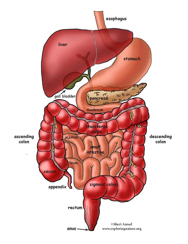
Digestion
In order for nutrients and minerals to be absorbed into blood , food we eat must be broken down into smaller molecules. The digestive system breaks down ingested food and liquid into smaller molecules our body can absorb. In turn, the nutrient-rich blood is then distributed throughout the body by the cardiovascular system.

Metabolism
Metabolism is a term that includes all chemical reactions that occur within the body. Metabolism is regulated by hormones secreted from (glands) the endocrine system.

Excretion
If the human body is to operate correctly, it must be able to get rid of waste and nonessential items. Several organ systems participate in the excretion of waste products. For instance, the urinary system disposes nitrogenous waste while the digestive system rids the body of indigestible food as feces.

Reproduction
In order to survive, all living organisms must reproduce. The reproductive system is responsible for producing offspring and is directly regulated by hormones of the endocrine system.

Growth
Humans must grow to survive. The scientific term for growth means “constructive activities must occur at a faster rate than destructive ones”.

Survival Needs

Survival needs include nutrients (food), oxygen, water and an appropriate atmosphere.

Nutrients
Nutrients contained in food and liquids contain chemical substances used for energy and cell building. Carbohydrates, vitamins, minerals, proteins, and fats are all vital in maintaining a healthy body. For instance, calcium helps make bones hard and vitamin D is needed in order to produce sufficient amounts of calcium.

Oxygen
Human cells can only survive for a few minutes without oxygen. Chemical reactions that release energy from foods are oxidative reactions and require oxygen. In fact, oxygen is so vital to the human body that it would only last a few minutes without it. The respiratory and cardiovascular systems work in conjunction to make oxygen available throughout the body.

Image result for drinking water

Water
The human body is 60-80% water. Water is the most abundant chemical substance in the body and provides the environment necessary for life. We obtain water through food and liquids and loose it through bodily excretions and evaporation (from the skin).

Appropriate atmospheric pressure
Breathing and gas exchange in the lungs depend on the right type of atmospheric pressure. For instance, on top of Mount Everest (at high altitude), gas exchange can be inadequate for the human body to survive.

Wednesday, January 25, 2017

Digestive System


The human gastrointestinal tract refers to the stomach and intestine, and sometimes to all the structures from the mouth to the anus.

The major organs of the human gastrointestinal system.

The major organs of the human gastrointestinal system are identified in this drawing. The upper gastrointestinal tract consists of the esophagus, stomach, and duodenum. The lower gastrointestinal tract includes most of the small intestine and all of the large intestine. According to some sources, it also includes the anus.

Upper Gastrointestinal Tract

The upper gastrointestinal tract consists of the esophagus, stomach, and duodenum. The exact demarcation between upper and lower can vary. Upon gross dissection, the duodenum may appear to be a unified organ, but it is often divided into two parts based upon function, arterial supply, or embryology.

The upper gastrointestinal tract includes the:

Esophagus, the fibromuscular tube that food passes through—aided by peristaltic contractions—the pharynx to the stomach.

Stomach, which secretes protein-digesting enzymes called proteases and strong acids to aid in food digestion, before sending the partially digested food to the small intestines.

Duodenum, the first section of the small intestine that may be the principal site for iron absorption.

Lower Gastrointestinal Tract

The lower gastrointestinal tract includes most of the small intestine and all of the large intestine. According to some sources, it also includes the anus.

Upper and lower gastrointestinal tract


The small intestine has three parts:

Duodenum: Here the digestive juices from the pancreas (digestive enzymes) and the gallbladder (bile) mix together. The digestive enzymes break down proteins and bile and emulsify fats into micelles. The duodenum contains Brunner's glands that produce bicarbonate, and pancreatic juice that contains bicarbonate to neutralize hydrochloric acid in the stomach.

Jejunum: This is the midsection of the intestine, connecting the duodenum to the ileum. It contains the plicae circulares and villi to increase the surface area of that part of the GI tract.

Ileum: This has villi, where all soluble molecules are absorbed into the blood ( through the capillaries and lacteals).

The large intestine has four parts:

1.Cecum, the vermiform appendix that is attached to the cecum.

2.Colon, which includes the ascending colon, transverse colon, descending colon, and sigmoid flexure. The main function of the colon is to absorb water, but it also contains bacteria that produce beneficial vitamins like vitamin K.

3.Rectum.

4.Anus.

The ligament of Treitz is sometimes used to divide the upper and lower GI tracts.


Image result for anatomy of digestive system pdf

Processes and Functions of the Digestive System

Digestion is necessary for absorbing nutrients from food and occurs through two processes: mechanical and chemical digestion.

The Digestive System

The proper functioning of the gastrointestinal (GI) tract is imperative for our well being and life-long health. A non-functioning or poorly-functioning GI tract can be the source of many chronic health problems that can interfere with your quality of life. 

Here is a look at the importance of two main functions of the digestive system: digestion and absorption.

Digestion

The gastrointestinal tract is responsible for the breakdown and absorption of the various foods and liquids needed to sustain life. Many different organs have essential roles in the digestion of food, from the mechanical breakdown of food by the teeth to the creation of bile (an emulsifier) by the liver. 

Bile production plays a important role in digestion: it is stored and concentrated in the gallbladder during fasting stages, and discharged to the small intestine. Pancreatic juices are excreted into the digestive system to break down complex molecules such as proteins and fats. 

Absorption

Absorption occurs in the small intestines, where nutrients directly enter the bloodstream.

Each component of the digestive system plays a special role in these complimentary processes. The structure of each component highlights the function of that particular organ, providing a seamless anatomy to keep our body fueled and healthy.

Components of the Digestive System

The digestive system is comprised of the alimentary canal, or the digestive tract, and other accessory organs that play a part in digestion—such as the liver, the gallbladder, and the pancreas. The alimentary canal and the GI tract are terms that are sometimes used interchangeably. 

The alimentary canal is the long tube that runs from the mouth (where the food enters) to the anus (where indigestible waste leaves). The organs in the alimentary canal include the mouth (the site of mastication), the esophagus, the stomach, the small and large intestines, the rectum, and the anus. From mouth to anus, the average adult digestive tract is about thirty feet (30') long.

Processes of Digestion

Food is the body's source of fuel. The nutrients in food give the body's cells the energy they need to operate. Before food can be used it has to be mechanically broken down into tiny pieces, then chemically broken down so nutrients can be absorbed. 

In humans, proteins need to be broken down into amino acids, starches into sugars, and fats into fatty acids and glycerol. This mechanical and chemical breakdown encompasses the process of digestion.

To recap these twin processes:

Mechanical digestion: Larger pieces of food get broken down into smaller pieces while being prepared for chemical digestion; this process starts in the mouth and continues into the stomach. 

Chemical digestion: Several different enzymes break down macromolecules into smaller molecules that can be absorbed. The process starts in the mouth and continues into the intestines. 

Moistening and Breakdown of Food

Digestion begins in the mouth. A brain reflex triggers the flow of saliva when we see or even think about food. Enzymes in saliva then begin the chemical breakdown of food; teeth aid in the mechanical breakdown of larger food particles.

Saliva moistens the food, while the teeth masticate the food and make it easier to swallow. To accomplish this moistening goal, the salivary glands produce an estimated three liters of saliva per day. 

Amylase, the digestive enzyme found in saliva, starts to break down starch into simple sugars before the food even leaves the mouth. The nervous pathway involved in salivary excretion requires stimulation of receptors in the mouth, sensory impulses to the brain stem, and parasympathetic impulses to salivary glands. Once food is moistened and rolled and ready to swallow, it is known as a bolus.

Swallowing and the Movement of Food

For swallowing to happen correctly a combination of 25 muscles must all work together at the same time. Swallowing occurs when the muscles in your tongue and mouth move the bolus into your pharynx. Bolus (from Latin bolus, ball)is a small rounded mass of a substance, especially of chewed food at the moment of swallowing.
"mucin holds the particles of food together in a ball or bolus" Mucin is a glycoprotein constituent of mucus.
"mucin is secreted by the salivary glands"

The pharynx, which is the passageway for food and air, is about five inches (5") long—a remarkably small space. A small flap of skin called the epiglottis closes over the pharynx to prevent food from entering the trachea, which would cause choking. Instead, food is pushed into the muscular tube called the esophagus. Waves of muscle movement, called peristalsis, move the bolus down to the stomach.

While in the digestive tract, the food is really passing through the body rather than being in the body. The smooth muscles of the tubular digestive organs move the food efficiently along as it is broken down into easily absorbed ions and molecules.

Large-scale Breakdown in the Stomach

Once the bolus reaches the stomach, gastric juices mix with the partially digested food and continue the breakdown process. The bolus is converted into a slimy material called chyme. 


Major digestive hormones

There are at least five major digestive hormones in the gut of mammals that help process food through chemical digestion in the gall bladder, duodenum, stomach, and pancrease. These hormones are cholecystokinin, gastric inhibitory polypeptide, motilin, secretin, and gastrin.

This (see above) is a drawing of the digestive system. This shows the five major digestive hormones in the gut of mammals that help process food through chemical digestion in the gall bladder, duodenum, stomach, and pancrease. These hormones are cholecystokinin, gastric inhibitory polypeptide, motilin, secretin, and gastrin.

The stomach is a muscular bag that maneuvers food particles, mixing highly acidic gastric juice and powerful digestive enzymes with the chyme to prepare for nutrient absorption in the small intestine. Stimulatory hormones such as gastrin and motilin help the stomach pump gastric juice and move chyme. The complex network of hormones eventually prepares chyme for entry into the duodenum, the first segment of the small intestine.

Absorption in the Small Intestine

During absorption, the nutrients that come from food (such as proteins, fats, carbohydrates, vitamins, and minerals) pass through the wall of the small intestine and into the bloodstream. In this way nutrients can be distributed throughout the rest of the body. The small intestine increases surface area for absorption through tiny interior projections, like small fingers, called villi.

Waste Compaction in the Large Intestine

In the large intestine there is resorption of water and absorption of certain minerals as feces are formed. Feces are the waste parts of the food that the body passes out through the anus.

Organs of the Digestive System

The organs of the digestive system can be divided into upper and lower digestive tracts. The upper digestive tract consists of the esophagus, stomach, and the small intestine; the lower tract includes all of the large intestine, the rectum, and anus.

The human body uses a variety of mental and physiological cues to initiate the process of digestion. Throughout our gastrointestinal (GI) tract, each organ serves a specific purpose to bring our food from the plate to a digestible substance from which nutrients can be extracted.

The Digestive Tube

Our digestive system is like a long tube, with different segments doing different jobs. The major organs within our digestive system can be split into two major segments of this tube: the upper gastrointestinal tract, and the lower gastrointestinal tract.

The Upper Gastrointestinal Tract

The upper gastrointestinal, or GI, tract is made up of three main parts: 

The esophagus.

The stomach.

The small intestine. 


The Lower Gastrointestinal Tract

The lower GI tract contains the remainder of the system: 

The large intestine.

The rectum.

The anus. 

The exact dividing line between upper and lower tracts can vary, depending on which medical specialist is examining the GI tract.

Food Breakdown and Absorption: The Upper GI Tract

When we take a bite of food, the food material gets chewed up and processed in the mouth, where saliva begins the process of chemical and mechanical breakdown. The chewing process is also known as mastication. 

When we mix up food with saliva, the resulting mushy wad is called a bolus. The bolus gets swallowed, and begins its journey through the upper gastrointestinal tract.

The Esophagus

The upper GI tract begins with the esophagus, the long muscular tube that carries food to the stomach. The throat cavity in which our esophagus originates is known as the pharynx. As we swallow, the bolus moves down our esophagus, from the pharynx to the stomach, through waves of muscle movement known as peristalsis. Next the bolus reaches the stomach itself. 

The Stomach

The stomach is a muscular, hollow bag that is an important part of the upper GI tract. Many organisms have a variety of stomach types, with many segments or even multiple stomachs. As humans, we have only one stomach. 

Here our bolus gets mixed with digestive acids, furthering breakdown of the bolus, and turning the bolus material into a slimy mess called chyme. The chyme moves on into the small intestine, where nutrients are absorbed.

The Small Intestine

The small intestine is an impressive digestive tube, spanning an average of 20 feet in length. The twists and turns of the small intestine, along with tiny interior projections known as villi, help to increase the surface area for nutrient absorption. 

This snaking tube is made up of three parts, in order from the stomach: 

The duodenum.

The jejunum. 

The ileum. 

As the chyme makes its way through each segment of the small intestine, pancreatic juices from the pancreas start to break down proteins. Soapy bile from the liver, stored in the gallbladder, gets squirted into the small intestine to help emulsify—or break apart—fats. 

Now thoroughly digested, with its nutrients absorbed along the path of the small intestine, what remains of our food gets passed into the lower GI tract.

Waste Compaction and Removal: The Lower Gastrointestinal Tract

The Large Intestine (Colon)

Following nutrient absorption, the food waste reaches the large intestine, or colon. The large intestine is responsible for compacting waste material, removing water, and producing feces—our solid-waste product. 

Accessory organs like the cecum and appendix, which are remnants of our evolutionary past, serve as special pockets at the beginning of the large intestine. The compacted and dried-out waste passes to the rectum, and out of the body through the anus. Healthy gut bacteria in the large intestine also help to metabolize our waste as it finishes its journey.


Enteric Nervous System

The enteric nervous system (ENS) is a subdivision of the autonomic nervous system (ANS) that directly controls the gastrointestinal system.

The gastrointestinal (GI) system has its own nervous system, the enteric nervous system (ENS). Neurogastroenterology is the study of the enteric nervous system, a subdivision of the autonomic nervous system (ANS) that directly controls the gastrointestinal system. The ENS is capable of autonomous functions such as the coordination of reflexes. 

Although it receives considerable innervation from the autonomic nervous system, it can and does operate independently of the brain and the spinal cord. The ENS consists of some 100 million neurons, one-thousandth of the number of neurons in the brain, and about one-tenth the number of neurons in the spinal cord. The enteric nervous system is embedded in the lining of the gastrointestinal system.

Ganglia of the ENS

The neurons of the ENS are collected into two types of ganglia: 

1.The myenteric (Auerbach's) plexus, located between the inner and outer layers of the muscularis externa.

2.The submucosal (Meissner's) plexus, located in the submucosa. 

The Myenteric Plexus 

The myenteric plexus is mainly organized as a longitudinal chains of neurons. When stimulated, this plexus increases the tone of the gut as well as the velocity and intensity of its contractions. This plexus is concerned with motility throughout the whole gut. Inhibition of the myenteric system helps to relax the sphincters—the muscular rings that control the flow of digested food or food waste. 

The Submucosal Plexus

The submucosal plexus is more involved with local conditions and controls local secretion and absorption, as well as local muscle movements. The mucosa and epithelial tissue associated with the submucosal plexus have sensory nerve endings that feed signals to both layers of the enteric plexus. These tissues also send information back to the sympathetic pre-vertebral ganglia, the spinal cord, and the brain stem.


Neural control of the gut.
An illustration of neural control of the gut wall by the autonomic nervous system and the enteric nervous system.


Function and Structure of the ENS

The enteric nervous system has been described as a second brain. There are several reasons for this. For instance, the enteric nervous system can operate autonomously. It normally communicates with the central nervous system (CNS) through the parasympathetic (e.g., via the vagus nerve) and sympathetic (e.g., via the prevertebral ganglia) nervous systems. However, vertebrate studies show that when the vagus nerve is severed, the enteric nervous system continues to function.

In vertebrates, the enteric nervous system includes efferent neurons, afferent neurons, and interneurons, all of which make the enteric nervous system capable of carrying reflexes and acting as an integrating center in the absence of CNS input. For instance, the sensory neurons report mechanical and chemical conditions, while the motor neurons control peristalsis and the churning of intestinal contents through the intestinal muscles. Other neurons control the secretion of enzymes. 

The enteric nervous system also makes use of more than 30 neurotransmitters, most of which are identical to the ones found in the CNS, such as acetylcholine, dopamine, and serotonin. More than 90% of the body's serotonin is in the gut, as well as about 50% of the body's dopamine, which is currently being studied to further our understanding of its utility in the brain.

The enteric nervous system has the capacity to alter its response depending on factors such as bulk and nutrient composition. In addition, the ENS contains support cells that are similar to the astroglia of the brain, as well as a diffusion barrier around the capillaries that surround the ganglia, which is similar to the blood–brain barrier of the cerebral blood vessels.

Regulation of ENS Function

The parasympathetic nervous system is able to stimulate the enteric nerves in order to increase enteric function. The parasympathetic enteric neurons function in defecation and provide a rich nerve supply to the sigmoid colon, the rectum, and the anus.

Conversely, stimulation of the enteric nerves by the sympathetic nervous system will inhibit enteric function and capabilities. Neurotransmitter secretion and direct inhibition of the enteric plexuses cause this stall in function. If the gut tract is irritated or distended, afferent nerves will send signals to the medulla of the brain for further processing.

Gastrointestinal Reflex Pathways

The digestive system functions via a system of long reflexes, short reflexes, and extrinsic reflexes from gastrointestinal (GI) peptides that work together.

Food in the Digestive System

The digestive system has a complex system of food movement and secretion regulation, which are vital for its proper function. Movement and secretion are regulated by long reflexes from the central nervous system (CNS), short reflexes from the enteric nervous system (ENS), and reflexes from the gastrointestinal system (GI) peptides that work in harmony with each other. 

In addition, there are three overarching reflexes that control the movement, digestion, and defecation of food and food waste: 

1.The enterogastric reflex.

2.The gastrocolic reflex.

3.The gastroileal reflex.

Long and Short Reflexes

Long reflexes to the digestive system involve a sensory neuron that sends information to the brain. This sensory information can come from within the digestive system, or from outside the body in the form of emotional response, danger, or a reaction to food. 

These alternative sensory responses from outside the digestive system are also known as feedforward reflexes. Emotional responses can also trigger GI responses, such as the butterflies in the stomach feeling when nervous. 

Control of the digestive system is also maintained by enteric nervous system (ENS), which can be thought of as a digestive brain that helps to regulate motility, secretion, and growth. The enteric nervous system can act as a fast, internal response to digestive stimuli. When this occurs, it is called a short reflex. 

Three Main Types of Gastrointestinal Reflex

1.The enterogastric reflex is stimulated by the presence of acid levels in the duodenum at a pH of 3–4 or in the stomach at a pH of 1.5. When this reflex is stimulated, the release of gastrin from G-cells in the antrum of the stomach is shut off. In turn, this inhibits gastric motility and the secretion of gastric acid (HCl). Enterogastric reflex activation causes decreased motility.

2.The gastrocolic reflex is the physiological reflex that controls the motility, or peristalsis, of the gastrointestinal tract. It involves an increase in motility of the colon in response to stretch in the stomach and the byproducts of digestion in the small intestine. Thus, this reflex is responsible for the urge to defecate following a meal. The small intestine also shows a similar motility response. The gastrocolic reflex also helps make room for food in the stomach.

3.The gastroileal reflex is a third type of gastrointestinal reflex. It works with the gastrocolic reflex to stimulate the urge to defecate. This urge is stimulated by the opening of the ileocecal valve and the movement of the digested contents from the ileum of the small intestine into the colon for compaction.


Peristalis
The gastrocolic reflex is one of a number of physiological reflexes that control the motility, or peristalsis, of the gastrointestinal tract.


GI Peptides that Contribute to Gastrointestinal Signals
GI peptides are signal molecules that are released into the blood by the GI cells themselves. They act on a variety of tissues that include the brain, the digestive accessory organs, and the GI tract. 

The effects range from excitatory or inhibitory effects on motility and secretion, to feelings of satiety or hunger when acting on the brain. These hormones fall into three major categories: 

1.The gastrin family.

2.The secretin family.

3.A third family that is composed of the hormones that do not fit into either of these two families.


Tuesday, August 21, 2012

THE NEW PARADIGM

THE NEW PARADIGM

"A new scientific truth is not usually presented in a way to convince its opponents. Rather, they die off, and a rising generation is familiarized with the truth from the start."-Max Planck

The new scientific truth and level of thinking about the human body that will empower people to become practitioners of preventive medicine for themselves is as follows: It is the solvent— the water content—that regulates all functions of the body, including the activity of all the solutes (the solids) that are dissolved in it. The disturbances in water metabolism of the body (the solvent metabolism) produces a variety of signals, 

indicating a "system" disturbance in the particular functions associated with the water supply and its rationed regulation.

Let me repeat: every function of the body is monitored and pegged to the efficient flow of water. 'Water distribution"is the only way of making sure that not only an adequate amount of water, but its transported elements (hormones,chemical messengers and nutrients) first reach the more vital organs. In turn, every organ that produces a substance to be made available to the rest of the body will only monitor its own rate and standards of production and release into the "flowing water," according to constantly changing quotas set by the brain. Once the water itself reaches the "drier" areas, it also exercises its many other most vital and missing physical and chemical regulatory actions.

Within this view, water intake and its priority distribution achieve paramount importance. The regulating neurotransmitter systems (histamine and its subordinate agents) become increasingly active during the regulation of water requirements of the body. Their action should not be continuously blocked by the use of medication. Their purpose should be understood and satisfied by drinking more water. I have made exactly the same statements to a body of scientists that had gathered from all over the world in Monte Carlo in 1989 for a conference on the topic of inflammation, analgesics, and immune modulators.


The new paradigm permits an incorporation of the "fourth dimension of time" into scientific research. It will facilitate an understanding of the damaging effect of an establishing dehydration that persists and continues to increase during any duration of time. It will make it possible to forecast the physiological events that will lead to disease states at some later years, including what at present appears as genetic disorders. It will transform the present"shot-in-the-dark, symptoms-treating" approach to the practice of medicine into a scientifically accurate medical art;it will make preventive forecasting possible. It will establish excellent health and reduce health care costs to individuals and to any society that fosters its spread. Since water shortage in different areas of the body will manifest varying symptoms, signals, and complications now labeled as diseases, people may think water could not be offered as a natural solution. Water cures so many diseases? No way! Speaking thus, they shut their minds to the new possibility of preventing and possibly even curing so many different "diseases" that are dehydration produced. It does not occur to them that the only remedy for conditions that come about when the body begins to get dehydrated is water and nothing else. A number of sample testimonials are published in different sections of this book to open the eyes of skeptics to the fact that the greatest health discovery of all times is that water is a natural medication for a variety of health conditions.

Water Regulation at Different Stages of Life

There are basically three stages to water regulation of the body in the different phases of life. One, the stage of life of a fetus in the uterus of the mother (left of B in Figure 1). Two, the phase of growth until full height and width is achieved (approximately between the ages of 18 to 25). Three, the phase of life from fully grown to the demise of the person. During the intrauterine stage of cell expansion, water for cell growth of the child has to be provided by the mother. However, the transmitter system for water intake seems to be produced by the fetal tissue, but registers its effect on the mother. The very first indicator for water needs of the fetus and the mother seems to be morning sickness during ' the early phase of pregnancy. Morning sickness of the pregnant mother-to-be is a thirst signal of both the fetus and the mother.