A bare foot was a sign of sensuousness, luxury, and pleasure.


 Ligaments are grouped together in cords, bands, or sheets. A sprain occurs when ligaments
covering a joint are torn or twisted. A sprained ankle is a partial tearing of the talofibular ligament that binds the bones of the lower leg to the bones of the foot. Ligaments heal slowly and they may not fully heal if they are completely torn apart.
C. sagittal: divides left from right.


3. The single plane motions of the foot:


4. Pronation and supination: There are two motions of the foot, pronation and supination, which include simultaneous movement in the frontal, sagittal, and transverse planes. These are termed tri-plane movements.
Pronation is a tri-plane motion consisting of simultaneous movements of abduction, dorsiflexion, eversion.


Supination is a tri-plane motion which combines the movements of adduction, plantarflexion, inversion.


It is difficult to clinically measure a tri-plane motion in the ankle at the subtalar joint (“STJ”).
Therefore, frontal plane motion is used as an index to measure tri-plane motion at the STJ. The number of degrees of inversion or eversion in the frontal plane signifies the amount of pronation and supination.
As the foot strikes the ground it immediately begins pronating to absorb shock and acts as a “mobile adaptor” (“loose bag of bones”) for variance in the terrain. It must then serve as a “rigid lever” to propel the body forward in locomotion. The latter occurs when the foot is supinated, as the foot structure becomes more rigid when supinated.
5. lateral and medial - Lateral means on the side away from the mid-line sagittal plane and medial means on the side closer to the mid-line sagittal plane.
6. dorsum and plantar surfaces - The dorsum is the top part of the foot. The plantar surface is the sole of the foot.
7. positions of the foot:
a. dorsiflexed and plantarflexed: In the normal foot, the reference point for a dorsiflexed or plantarflexed position is a transverse plane which runs through the heel. If the foot is positioned below this transverse plane, it is said to be plantarflexed; above this transverse plane, it is said to be dorsiflexed.
b. Everted and inverted: A foot or part of a foot is said to be inverted when it is tilted parallel to the frontal plane so that the plantar surface of the foot or part of the foot faces toward the midline of the body. A foot or part of the foot is said to be everted when it is tilted parallel to a frontal plane so that the plantar surface faces away from the midline of the body.
c. abducted and adducted: The two transverse plane positions of the foot are abducted and adducted. The reference point is the mid-line sagittal plane.
8. Fixed structural positions of the foot can occur due to the inherent structure of bone, ligament, etc. of a particular foot.
a. Adductus and Abductus: Adductus denotes a fixed structural position in which the foot is held in an adducted position in the transverse plane. Abductus denotes a
fixed structural position in which the foot is held in an abducted position in the transverse plane.
b. Varus and Valgus: the two frontal plane fixed structural positions which the foot may assume relative to the inverted and everted positions. The fixed structural position in which the foot or part of the foot appears inverted is classified as varus. The fixed structural position in which the foot or part of the foot appears everted is classified as valgus.
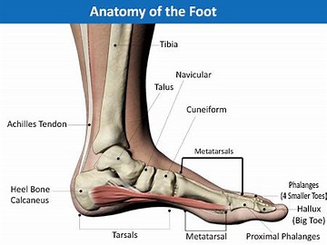
Basic Anatomy of the Foot
The foot is a perfect marriage of form and function. The foot contains 26 bones, 2 sesamoid
bones, 33 joints, 19 muscles and 107 ligaments.
The Musculoskeletal System
The skeletal system consists of bone, which is the hard substance that forms the framework of the body. Ligaments tie the bones together to form joints. Each bone and each joint has a name. The bone of the thigh is called the femur, the bones of the leg are called the tibia and fibula, and the joint between these bones is called the knee joint. The skeleton and all its parts are moved by muscles and tendons. Bones, ligaments, muscles, and tendons are the tissues of the locomotor or musculoskeletal system of the body.
Bones consist of two kinds of tissue:
1. compact tissue: the hard, outside part of a bone.
2. cancellous tissue: the spongy part on the inside.
Bones are covered with fibrous membrane called periosteum. The periosteum contains nerve fibres and transmits pain sensation if inflamed or torn away from the underlying bone.
The muscular system consists of muscle, which is the tough, elastic tissue that makes body parts move. The human body has more than 600 major muscles.
Skeletal muscles hold the bones of the skeleton together and make the body move.
Skeletal muscles vary greatly in size, depending on their function. For example, eye muscles are small and fairly weak, but thigh muscles are large and strong.
The ends of most skeletal muscles are attached to bones by a tough, flexible connective tissue called tendon. The origin of the muscle is the proximal end that is attached to bone that does not move when the muscle contracts (draws together). The distal end, the insertion, is attached to a bone that moves when the muscle contracts.
Skeletal muscles act in pairs called flexors and extensors.
1. flexor: bends a joint and decreases joint angle.
2. extensor: does the opposite, and moves a limb away from the body.
For example, the hamstring muscle at the back of the femur is a knee flexor. When it contracts, the knee bends and the leg moves toward the hip. The quadriceps muscle at the front of the femur is an extensor. When it contracts, the knee straightens and the leg moves away from the hip. At the same time, the hamstring relaxes so the quadriceps can pull the limb back to its original length.
Joints and Ligaments
A joint is the place that two or more bones meet. This is also called an articulation.
3
Freely-mobile joints such as the elbow and knee contain a synovial cavity. This cavity is lined inside by synovial membrane, which produces joint fluid (“synovial fluid”). The fluid reduces the friction of the moving bones. The synovial membrane is surrounded by a fibrous joint capsule which, in key places, is reinforced by ligaments.
Joints are protected from wear and tear in several ways. A smooth layer of cartilage (gristle) covers the end of bones that move over one another. The elasticity of cartilage breaks the force of sudden shocks and also, the smooth quality of the cartilage makes a joint move easily. In addition to cartilage, the synovial fluid keeps the joints moist and lubricated.
Bones are held together at the joint by strong ligaments that attach above and below the joint. The “joint capsule” encircles the joint and seals it to maintain the synovial fluid inside the capsule. This creates the “synovial cavity” (see diagram).
Medial view of left knee
A ligament is a fibrous tissue that holds organs of the body in place and fastens bones together.
covering a joint are torn or twisted. A sprained ankle is a partial tearing of the talofibular ligament that binds the bones of the lower leg to the bones of the foot. Ligaments heal slowly and they may not fully heal if they are completely torn apart.
Fascia is a broad connective tissue band serving a stabilization and supportive function (e.g. iliotibial band, plantar fascia).
Basic Anatomical Terms
There is a “language” used by medical practitioners of all kinds (podiatrists, chiropractors, orthopaedic surgeons, etc.). When discussing the body and its ailments, the basic terms you will need to know are:
1. proximal and distal: “proximal” means closer to the heart and “distal” means further away from the heart. Thus, each toe has three bones: the proximal phalanx, the middle phalanx, and the distal phalanx (except the big toe, which has two bones).
2. The three anatomical planes: transverse, frontal and sagittal – there are three planes that divide the body and are used as points of reference.
A. transverse: divides top and bottom.
B. frontal: divides front and back.
C. sagittal: divides left from right.
Transverse, Frontal and Sagittal planes.
Three Anatomical Planes of the Foot
When dealing with the foot, the midline is relative to the foot itself. Therefore, the midline of the
foot divides the 2nd and 3rd toe.3. The single plane motions of the foot:
A. Abduction and adduction: These movements occur in the transverse plane. The foot abducts when it rotates laterally (i.e. away from the centre). It adducts when it rotates medially (i.e. towards the centre.)
Abduction Neutral Adduction
Single transverse plane motion of the foot.
B. Inversion and eversion: These movements occur in the frontal plane. The foot inverts when it rotates inward and upward (the sole toward the midline), and everts when it rotates outward and upward (the sole away from the midline).
Eversion Neutral Position Inversion
Single frontal plane motion of the foot
Inversion and eversion of the foot (right foot).
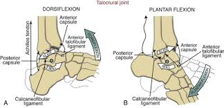
C. Plantarflexion and dorsiflexion: These movements occur in the sagittal plane. The foot plantar flexes when it moves downwards away from the tibia and dorsiflexes when it moves upwards toward the tibia.
Pronation is a tri-plane motion consisting of simultaneous movements of abduction, dorsiflexion, eversion.
Supination is a tri-plane motion which combines the movements of adduction, plantarflexion, inversion.
Therefore, frontal plane motion is used as an index to measure tri-plane motion at the STJ. The number of degrees of inversion or eversion in the frontal plane signifies the amount of pronation and supination.
As the foot strikes the ground it immediately begins pronating to absorb shock and acts as a “mobile adaptor” (“loose bag of bones”) for variance in the terrain. It must then serve as a “rigid lever” to propel the body forward in locomotion. The latter occurs when the foot is supinated, as the foot structure becomes more rigid when supinated.
5. lateral and medial - Lateral means on the side away from the mid-line sagittal plane and medial means on the side closer to the mid-line sagittal plane.
6. dorsum and plantar surfaces - The dorsum is the top part of the foot. The plantar surface is the sole of the foot.
7. positions of the foot:
a. dorsiflexed and plantarflexed: In the normal foot, the reference point for a dorsiflexed or plantarflexed position is a transverse plane which runs through the heel. If the foot is positioned below this transverse plane, it is said to be plantarflexed; above this transverse plane, it is said to be dorsiflexed.
b. Everted and inverted: A foot or part of a foot is said to be inverted when it is tilted parallel to the frontal plane so that the plantar surface of the foot or part of the foot faces toward the midline of the body. A foot or part of the foot is said to be everted when it is tilted parallel to a frontal plane so that the plantar surface faces away from the midline of the body.
c. abducted and adducted: The two transverse plane positions of the foot are abducted and adducted. The reference point is the mid-line sagittal plane.
8. Fixed structural positions of the foot can occur due to the inherent structure of bone, ligament, etc. of a particular foot.
a. Adductus and Abductus: Adductus denotes a fixed structural position in which the foot is held in an adducted position in the transverse plane. Abductus denotes a
fixed structural position in which the foot is held in an abducted position in the transverse plane.
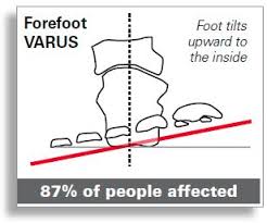
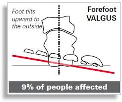
b. Varus and Valgus: the two frontal plane fixed structural positions which the foot may assume relative to the inverted and everted positions. The fixed structural position in which the foot or part of the foot appears inverted is classified as varus. The fixed structural position in which the foot or part of the foot appears everted is classified as valgus.

No comments:
Post a Comment