Showing posts with label pregnancy. Show all posts

Sunday, October 15, 2017

EXCUSE # 18: I'M PREGNANT

(OR PLANNING TO BE PREGNANT)

The good news is that going barefoot adds additional benefits for future mommies to be during pregnancy. Swollen feet are common during pregnancy, and many women find themselves shedding their shoes anyway. Consider these barefoot walking benefits of truly going barefoot:

Minimizes swollen feet. One of the big considerations for pregnant women is circulation. It's known that women's feet get bigger during pregnancy, and a large part of this is fluid gain, often caused by not working our muscles. However, fluid gain goes down as you use muscles more. When you go barefoot your feet have to wok more. That's a great thing when pregnant, because a working foot is like a fluid pump, bringing fluid up out of the foot and the lower leg and delivering it back to the heart.
Image result for pregnant leg secondary heart


Improves overall circulation. The increased circulation doesn't just end with the feet. Your lower legs also do more work when you're barefoot, particularly as you shift your weight toward your forefoot. This increased circulation acts as a fluid pump and helps keep your legs healthier, happier, and stronger. 
Image result for pregnant leg secondary fluid pump heart

Builds balance for less wobbling. One of the greatest benefits of going barefoot is that your feet are closer to the ground, which brings more stability during a time when your center of gravity is shifting. You can also feel the ground beneath your feet, which helps you reawaken and work the stabilizing muscle groups from your  feet to your legs, hips, and up into your core, improving your balance for increased maneuverability and lower chances of falling.

Strengthens core muscles for delivery day. If you think of the human body as a series of interconnected links of a chain, each one connected to the next, you start to get a real sense of the benefits of going barefoot when pregnant. After your legs, the next link in the chain is your core. Now, a stronger core isn't just about the stomach; it includes all the muscles from the pelvis on up. In essence, when you've fully engaged your core, you're doing somewhat of a Kegel exercise, just to hold yourself in place ㅡ muscles essential for an easier delivery day. 

Develops a healthier back for a happier mommy. 
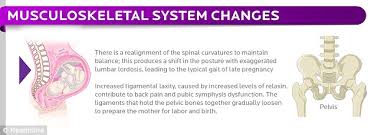
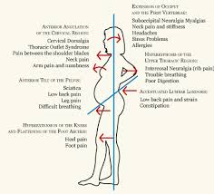
Keeping the back healthy and happy during pregnancy is often a big challenge. After all, you're carrying a growing heavy weight in front, and your back often arches forward.
Image result for pregnant woman back arches forward

Image result for pregnant woman back arches forward

Image result for pregnant woman back arches forward

And by working all of the muscles from your feet on up, you assist the circulation of your cerebrospinal fluid, in essence lubricating your spine and keeping it loose. 

Image result for lubricating your spine

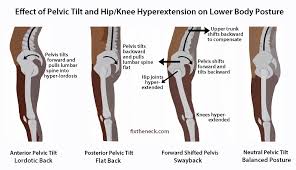
Stand with better posture for more comfort. Since you are working right up the chain and have better body awareness, better circulation, and a stronger core, you also end up with better posture, because you can feel what is going on beneath you ㅡ especially when there comes a time you can't see your feet. 
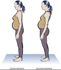
Image result for pregnant woman back arches forward

Image result for pregnant woman back arches forward

Image result for pregnant woman back arches forward

If you were  already walking barefoot before pregnancy, congratulations ㅡ you've got a leg up here. Keep doing what you're doing if your doctor agrees, and it will serve you well. But if you're already pregnant and would like to experience the benefits of being pregnant and going barefoot, always trust your doctor here, and when in doubt, be safe and ask.


Image result for pregnant woman barefoot walking

Image result for pregnant woman barefoot walking

Image result for pregnant woman barefoot walking

Image result for pregnant woman barefoot walking

Image result for pregnant woman barefoot walking

Image result for pregnant woman barefoot walking

Image result for pregnant woman barefoot walking

Image result for pregnant woman barefoot walking

Sunday, January 8, 2017

The True Power of Water: Healing And Discovering Ourselves

The True Power of Water: Healing and Discovering Ourselves
By Masaru Emoto, Noriko Hosoyamada

Hado Consultation for a Baby with Congenital Heart Disease Many people have come to see me for consultation. The reason I took on the following case is ...found in page 85 onwards, (Click Here) to discover the story .

The True Power of Water
By Masaru Emoto

Water is sensitive to a subtle form of energy called hado. It is this form of energy that affects the quality of water and the shape in which water crystals form.

In my previous book, The Hidden Messages in Water , the word hado was translated as ‘wave fluctuation'. In The True Power of Water, I use hado to mean all the subtle energy that exists in the universe.

All existing things have vibrations, or hado. This energy is often positive or negative and is easily transmitted to other existing things. The thought ‘You fool’ carries its own hado, which water absorbs and displays as deformed crystals when frozen. On the other hand, when water has been exposed to positive thoughts, beautiful crystals are formed that reflect the positive hado. Hado, as you can see, is integrally woven into the implications of water's response to information.

Hado medicine

Some time ago I was introduced to a type of radionics device [not available any longer] capable of measuring various vibrations of the body at the cellular level. I developed an affinity for the device and became able to use it beyond the device's intended design, namely, to measure hado. My experience with the hado device led me to realise water's ability to receive information. It also led the way to my study of hado medicine, an alternative medical practice that researchers started to study after having felt the limitations of conventional Western medicine.

Although having a device was helpful in my study of hado medicine, I came to believe that humans are capable of feeling and sending hado as well as the device can measure it, if not better. For example, healers and counsellors who can help their patients are considered to have the ability to send good vibrations to correct their patients' abnormal vibrational patterns.

The fundamental principles of hado medicine are vibration and resonance. When the cellular vibrations in different parts of the body are disturbed for various reasons, our body can make a wrong turn. When this situation occurs, a new external vibration can be given to the disturbed cell so as to resonate with it; thus, its intrinsic vibration is restored. This is hado medicine in a nutshell. How can the vibration be corrected?

A hado is a wave; it has a wave shape of peaks and valleys. When the shape of a wave opposite to the original one – valleys for peaks, and peaks for valleys – is used, the wave can be straightened. By overlaying a wave with another wave in this manner, its characteristics can be cancelled. Hado medicine utilizes this concept to restore the patient's health by sending the hado that can cancel his/her unfavourable hado. Water is an integral component, as you'll see.

To help a person treat himself or herself with hado medicine, we examine the individual's hado using the hado measuring device. After understanding the disturbances to his or her own vibration, we prepare the water on which the information to correct the vibration was transferred through the hado measuring device. The hado water created in this manner penetrates into the molecules, atoms, and subatomic particles that make up the person's body and stops the disturbances of this vibration. By drinking this hado water, the individual could correct the disturbed vibration.

How emotions affect the body

Through my studies, I noticed many common characteristics that diseased people share and discovered the close correlation between people's emotions and affected parts of their body.

In 1995, I conducted hado examinations on one hundred people. I measured their hado regarding the most commonly shared emotions (thirty-eight traits including stress, worry, pressure, irritability, perplexity and excess fear) and then checked which part of their body resonated the most with each emotion.

I found, for example, that those who feel stress tend to have problems with their intestines. Worries are often expressed as problems in the cervical nerves, irritabilities in the parasympathetic division of the autonomic nervous system, excess fear in the kidneys and anxiety in the stomach.

You might want to think about your present emotions and physical conditions. Do they agree with these findings? It's true that ‘Worry is often the cause of illness’. When your emotional conditions improve, your illness often moves toward recovery.

Try saying an antonym(a word opposite in meaning to another ;e.g. bad and good ) of the unfavourable emotion that you are feeling. If you are feeling ‘stress’, the antonym is ‘relaxation', and for ‘irritability', it is ‘calm'. The most basic solution to correct your hado is to use the opposite word. To take this approach further, write the antonyms on a sheet of paper and show it to water. The information is given to the water; in other words, the hado of the positive trait is transmitted to the water. Then, when you drink it, you are in effect practising hado medicine.


From The True Power of Water, by Masaru Emoto


Factors Contributing to Congenital Heart Disease

What causes congenital heart disease?

A congenital heart defect (CHD) is a heart problem which is present at birth. It is caused by abnormal formation of the heart during fetal development. In most cases, when a baby is born with congenital heart disease, there is no known reason for it. Scientists know that some types of congenital heart defects can be related to an abnormality in the number of an infant's chromosomes, single gene defects, or environmental factors. In most cases, there is no identifiable cause for the heart defect, and they are generally considered to be caused by multifactorial inheritance.

Multifactorial inheritance means that "many factors" (multifactorial) are involved in causing a birth defect. The factors are usually both genetic and environmental, where a combination of genes from both parents, in addition to unknown environmental factors, combine to produce the trait or condition.

Maternal factors and CHD

Most mothers of babies born with congenital heart disease will look critically at their own behaviors during pregnancy to try to find a cause for their child's illness. It is important to remember that most cases of congenital heart disease have no known cause. However, some types of congenital heart defects are known to occur more often when the mother comes in contact with certain substances during the first few weeks of pregnancy, while the baby's heart is developing. Some maternal illnesses and medications taken for these illnesses have been shown to affect the heart's development. Other illnesses or medications seem to have no impact on the baby's heart. Always consult your health care provider or obstetrician for more information.

Women who have seizure disorders and need to take antiseizure medications may have a higher risk for having a child with congenital heart disease, as do women who take lithium to treat depression. Mothers who have phenylketonuria (PKU) who do not adhere to the special diet necessary to manage the disease during pregnancy have a higher risk of having a child with congenital heart disease. Also, women with insulin-dependent diabetes (particularly if the diabetes is not well-controlled) or lupus may have a higher risk of having a child with heart defects. Counseling is important for women with these chronic illnesses before becoming pregnant.

Rubella, a virus that most people in the United States were immunized against when they received the MMR vaccine, is known to cause birth defects. A woman who has never had rubella nor been vaccinated against the disease should consult her health care provider before becoming pregnant. A mother who contracts rubella during her pregnancy has a very significant chance of having a baby with birth defects, including congenital heart disease.

Always consult your health care provider before taking any medications while you are pregnant.

Family history and CHD

In the general population, about 1% of all children are born with congenital heart disease. However, the risk increases when either parent has CHD, or when another sibling was born with CHD.

Some heart defects are considered to have autosomal-dominant inheritance. This means that a parent with the defect has a 50% chance, with each pregnancy, to have a child with the same heart defect, and males and females are equally affected. Similarly, there is also a 50% chance that a baby will not be affected.

Consultation with a genetic counselor or genetic specialist is encouraged for women with congenital heart disease before becoming pregnant. In families with CHD either in the parents or prior children , fetal echocardiography can be done in the second trimester, at about 18 to 22 weeks of pregnancy, to look for the presence of major heart defects in the fetus.

Chromosome abnormalities and CHD
Chromosomes are the structures in your cells that contain your genes. Genes contain the code for your traits such as eye color and blood type. Usually, there are 46 chromosomes in each cell of the body. Having too many or too few chromosomes results in health problems and birth defects. Structural defects of the chromosomes, where a piece of a chromosome is missing, or present in duplicate, also cause health problems.

Problems with chromosomes that result in genetic syndromes, such as Down syndrome, often result in a higher incidence of infant heart malformations. Among infants with chromosome abnormalities, around 30% will have a heart defect. 

There are a number of chromosome abnormalities associated with congenital heart defects. Some of these include the following:

Down syndrome (trisomy 21)

Trisomy 18 and trisomy 13

Turner's syndrome

Cri-du-chat syndrome

Wolf-Hirshhorn syndrome

DiGeorge syndrome (22q11)

Chromosome analysis can be done from a small blood sample to look for a chromosome abnormality in a child with a congenital heart defect.

Single gene defects
There are an estimated 70,000 genes contained on the 46 chromosomes in each cell of the body. Genes come in pairs, one of which is inherited from the mother, the other from the father. Genes not only help to determine our individual traits, but also may be responsible for health problems when gene alterations (mutations) are present. When a single gene is mutated, a number of health problems may occur in a person. when several health problems occur from one genetic cause that is referred to as a syndrome. Some of the genetic syndromes associated with a higher incidence of heart defects include, but are not limited to, the following:

Marfan syndrome

Smith-Lemli-Opitz syndrome

Ellis-van Creveld syndrome

Holt-Oram syndrome

Noonan syndrome

Mucopolysaccharidoses

Other genetic syndromes that are not due to a single gene defect, but are associated with CHD, include Goldenhar syndrome (hemifacial microsomia), William's syndrome, and the VACTERL association (tracheal and esophageal malformations associated with vertebral, anorectal, cardiac, renal, and limb abnormalities.)

When a child is born with a congenital heart defect, if there is a suspicion that the child has some type of genetic syndrome, a doctor who specializes in genetics (called a clinical geneticist) may be asked to evaluate your child.


If a child has been diagnosed with a chromosomal or other genetic abnormality, genetic counseling is helpful to determine the risk for heart defects occurring in future children.

WATER REVIVES LIFE FORCE
(PAGE 79)
Hado Consultation for a Girl with Acute Myclocytic Leukemia.

On February 24, 1996, I was asked to work with a girl, then fourteen years old, who had been recently attacked by leukemia. It may be a little bit lengthy but I would like to show you the process of her recovery based on her records. 

Wednesday, December 21, 2016

Do Not 'Kid' Your Kidneys

Kidney Function Blood Test

Routine kidney function is one of the most commonly performed blood tests.

The kidneys

The kidneys regulate the amount of water and salts that we have in our bodies. They do this by filtering the blood through millions of structures called nephrons. The kidneys also pass out certain waste products from the body. Urine is made up of the excess water, salts and waste products passed out by the kidneys down to the bladder.


Routine blood test of kidney function.
The usual blood test which checks that the kidneys are working properly measures the level of urea, creatinine and certain dissolved salts.

Urea is a waste product formed from the breakdown of proteins. Urea is usually passed out in the urine. A high blood level of urea ('uraemia') indicates that the kidneys may not be working properly, or that you have a low body water content (are dehydrated).

Creatinine is a waste product made by the muscles. Creatinine passes into the bloodstream, and is usually passed out in urine. A high blood level of creatinine indicates that the kidneys may not be working properly. Creatinine is usually a more accurate marker of kidney function than urea. The effect of muscle mass needs to be taken into account. A person with a lot of muscle and little fat on their body is likely to have a higher creatinine than a person who has a lot of fat and little muscle.

Estimated glomerular filtration rate (eGFR) provides a guide to kidney function. Although the level of creatinine in the blood is a useful guide to kidney function, the eGFR is a more accurate measure. Blood creatinine can be used to estimate the eGFR using age, sex and race. This is often calculated by computer and reported with the creatinine blood test. The normal value for eGFR is 90-120 ml/min. An eGFR below 60 ml/min suggests that some kidney damage has occurred. The value becomes lower with increasing severity of kidney damage.

Dissolved salts that are routinely measured are sodium, potassium, chloride and bicarbonate. They are sometimes referred to as 'electrolytes'. Abnormal blood levels of any of these may be due to a kidney problem. (Some other conditions may also alter the salt balance in the blood.)

[[Estimated Glomerular Filtration Rate
Various different diseases, conditions and medicines can affect the function of the kidneys. The estimated glomerular filtration rate (eGFR) does not diagnose any kidney disease but is a test to
assess how well your kidneys are working. A reading of greater than 90 ml/min/1.73m2 is normal.

What is the estimated glomerular filtration rate (eGFR)?

The eGFR is a test that is used to assess how well your kidneys are working. The test estimates the volume of
blood that is filtered by your kidneys over a given period of time. The test is called the estimated glomerular
filtration rate because the glomeruli are the tiny filters in the kidneys. If these filters do not do their job properly
then the kidney is said to have reduced or impaired kidney function.

The eGFR test involves a blood test which measures a chemical called creatinine. Creatinine is a breakdown
product of muscle. Creatinine is normally cleared from the blood by the kidneys. If your kidneys are not working
properly, the level of creatinine in the blood goes up.

The eGFR is then calculated from your age, sex and blood creatinine level. An adjustment to the calculation is
needed for people with African-Caribbean origin.

Stages of kidney function

The level of kidney function is divided into five stages:
Stage of Chronic Kidney Disease (eGFR;ml/min/1.73m)

Stage 1: the eGFR shows normal kidney function but you are already known to have some kidney damage or disease. For example, you may have some protein or blood in your urine, an abnormality of your kidney, kidney inflammation, etc.
(90 or more)

Stage 2: mildly reduced kidney function AND you are already known to have some kidney damage or disease. People with an eGFR of 60-89 without any known kidney damage or disease are not considered to have chronic kidney disease (CKD).(60 to 89)

Stage 3: moderately reduced kidney function. (With or without a known kidney disease. For example, an elderly person with ageing kidneys may have reduced kidney function without a specific known kidney disease.) (45 to 59)-(3A); (30 to 44)-(3B)

Stage 4: severely reduced kidney function. (With or without known kidney disease.)  (15 to 29)

Stage 5: very severely reduced kidney function. This is sometimes called end-stage kidney failure or established renal failure.
(Less than 15)

Note: it is not possible to assess the estimated glomerular filtration rate (eGFR) accurately in people with abnormal amounts of muscle and in people who have conditions that can affect the level of creatinine. This includes:

>People with muscle wasting conditions.

>People who have had an amputation of an arm or leg.

>Malnourished people.

>Pregnancy.

>People with acute kidney failure.

>People with a lot of fluid retention (edema).

>Children.]]

Who has a blood test of kidney function?

 Routine kidney function is one of the most commonly performed blood tests. It may be done:

>As part of a general health assessment.

>If you have suspected low body water content (dehydration), when the urea level increases.

>If you have suspected kidney failure.(Read below, Acute Kidney Injury)  The higher the blood levels of urea and creatinine, the less well the kidneys are working. The level of creatinine is usually used as a marker as to the severity of kidney failure. Creatinine in itself is not harmful but a high level indicates that the kidneys are not working properly. So, many other waste products will not be cleared out of the bloodstream. You normally need treatment with dialysis if the level of creatinine goes higher than a certain value.


>Before and after starting treatment with certain medicines. Some medicines occasionally cause kidney damage as a side-effect. Therefore, kidney function is often checked before and after starting treatment with certain medicines.

Other tests of kidney function


The routine kidney blood test is a general marker of kidney function. If the blood test is abnormal it cannot say what is causing the kidney problem. Therefore, if you have an abnormal result you may need further tests to find the cause of a kidney problem. For example: urine tests, other blood tests, scans, X-rays, kidney biopsy, etc.

Acute Kidney Injury

Acute kidney injury is a condition where your kidneys suddenly stop working properly. Usually this happens when you are unwell from another illness. If you have acute kidney injury you are often already ill in hospital. If not, you will usually be sent to hospital to be looked after. The earlier acute kidney injury is picked up, and the more carefully you are monitored, the better the chance of your kidneys recovering.

Acute kidney injury used to be called acute renal failure.


Understanding kidneys: where they are and what they do

The two kidneys lie to the sides of the upper part of the tummy (abdomen), behind the intestines, and either side of the spine. Each kidney is about the size of a large orange but bean-shaped.


A large artery - the renal artery - takes blood to each kidney. The artery divides into many tiny blood vessels (capillaries) throughout the kidney. In the outer part of the kidneys tiny blood vessels cluster together to form structures called glomeruli.

Each glomerulus is like a filter. The structure of the glomerulus allows waste products and some water and salt to pass from the blood into a tiny channel called a tubule. The liquid that remains at the end of each tubule is called urine. The urine then passes down a tube called a ureter which goes from each kidney to the bladder. Urine is stored in the bladder until it is passed out when we go to the toilet.

The main functions of the kidneys are to:

>Filter out waste products from the bloodstream, to be passed out in the urine.

>Help control blood pressure - partly by the amount of water passed out of the body as urine and partly by making hormones which are involved in blood pressure control.

>Make a hormone called erythropoietin, which stimulates the bone marrow to make red blood cells. This is needed to prevent anaemia.

>Control the amount of fluid in the body.

>Help keep various salts and chemicals in the blood at the right level.

What is acute kidney injury (AKI)?

If you have AKI your kidneys suddenly stop working as well as they should. This means the fluid, salts and chemicals in your body can rise or fall to abnormal levels and make you very ill. If there is too much acid, or there are too many salts, in your body your other organs can be affected. For example, your heart, lungs, brain, muscles, eyes, etc, can stop working properly. If the kidneys aren't getting rid of enough fluid, your body can become overloaded with it. This can cause swelling in your legs, or make it difficult to breathe.

It is different from chronic kidney disease (CKD) which is a much more gradual process which can happen over many years.

How common is acute kidney injury (AKI)?

In people who are unwell in hospital, AKI is common. Nearly as many as 1 in 5 people in hospital develop AKI. It is even more common in patients who are extremely unwell in intensive care units.

Who develops acute kidney injury (AKI)?

AKI is more common over the age of 65. People who have another serious illness and are in hospital are more likely to develop it. It may happen to people who are having operations, especially if they are older. It may happen to some people who are having a special X-ray with a dye (contrast) which contains iodine.

Some things make you more likely to develop AKI in these situations - for example:

>Being older.

>Having another illness like diabetes, heart failure or liver disease.

>Being dependent on a carer for fluids.

>Having a bad infection (for example, from a urinary infection, chest infection or skin infection).

>Some medicines - for example:
   -Non-steroidal anti-inflammatory drugs (NSAIDs), such as ibuprofen, diclofenac or naproxen.
    -Some blood pressure pills that work on the kidney.
    -Some antibiotics called aminoglycosides (such as gentamicin).
    -'Water tablets' (diuretics).

>Lacking in fluid in the body (being dehydrated).

>Another problem with your kidneys, such as CKD.

>Having had AKI before.

Children can develop AKI too. This is more likely if they have:

>Severe diarrhoea.

>A cancer of the blood cells.

>To rely on a parent or carer for fluids (more likely to become dehydrated).

>Low blood pressure.

>Another kidney illness such as nephritis.


>A severe infection.

What causes acute kidney injury (AKI)?


There are many things that can cause the kidneys to stop working properly. Sometimes it happens due to more than one cause.

Causes include:

>Severe infections.

>Blockage in the urinary system.

>Lack of fluid in the body (dehydration).

>Heavy bleeding (haemorrhage).

>Low blood pressure.

>Some medicines (see previous section for medicines that can harm the kidneys).

>Kidney diseases.


>The dye (contrast) that is used for some kinds of scans and X-rays.

What are the symptoms of acute kidney injury (AKI)?

There may be no specific symptoms of AKI. Often you already have symptoms of the illness that has caused AKI.

Symptoms that may happen include:

>Passing less urine than usual.

>Feeling sick or being sick (vomiting).

>Poor appetite.

>Breathlessness.

>Swelling in the legs or other parts of the body.

How is acute kidney injury (AKI) diagnosed?

Your doctor would test for AKI with urine tests and blood tests. Your urine will be measured to see how much you are passing. In most people with AKI, this is less than normal. It will also be tested with a 'dipstick' to check for protein, blood cells and sugar, amongst other things. This will help work out the cause. You will have a blood test for a substance called creatinine. This is an indicator for how well the kidneys are working. Higher levels mean that the kidneys aren't getting rid of waste products as effectively as they should.

You may need an ultrasound scan to look for blockages in the urinary system. Ultrasound scans use sound waves to create a picture of the kidneys and urinary system.

You may also need other tests to try to find the cause for AKI. These might include other blood tests, scans or X-rays.

How is acute kidney injury (AKI) treated?

There isn't a specific medicine to treat AKI. The treatment will depend on the cause of AKI and the severity of the AKI.

The treatment is to carefully look after all the things the kidneys usually do. You will normally be in hospital and have blood and urine tests regularly. You may need extra or special fluid through a drip. You may be on a restricted diet to help get the balance of proteins and salts right. Any medicines which might be harming your kidneys would be stopped. The cause of the problem would be treated if this is possible. (For example, you might need antibiotics for an infection or surgery for any blockage if these were the cause.)

Your doctor will keep a close eye on the balance of minerals and salts in your body. You will have regular tests for potassium, sodium, calcium, sugar (glucose), phosphate and creatinine. You may need treatment with medicines or through a drip if the levels are not right.

Sometimes you need to have dialysis if medicines and fluids aren't working and you are becoming more ill. Dialysis is a procedure where your blood is passed through a machine to remove the waste products (as your kidneys would do if they were working). There are different ways this is done. How often it is done and how long each session lasts will depend on the way it is done. You would be referred to a kidney specialist or an intensive care specialist for this. In most people this can be stopped once the kidneys are working properly again.


These decisions would be made after discussion with you and explaining the options to you (and/or your relatives or carers where appropriate).

What is the outlook (prognosis)?

The outlook varies hugely. It depends on how bad kidney function becomes, who is looking after you, the cause of the problem and how ill you were to begin with.

The National Institute for Health and Care Excellence (NICE) has produced guidelines to try to cut the numbers of people who develop acute kidney injury (AKI) and who die because of it. The outlook is better if it is picked up earlier.

Many people who recover from AKI have normal kidney function afterwards. However, some go on to develop chronic kidney disease (CKD). Some need to keep having dialysis. If you have had AKI once, you are more likely to have it again in the future.

How can acute kidney injury (AKI) be prevented?

Prevention is certainly better than cure for AKI. Patients in hospital should be tested for AKI regularly. This is done by frequent blood tests and measuring how much urine they produce (and weight for children). People having scans or X-rays with a dye (contrast) should be checked to see how likely they are to develop AKI. If they are at risk, and if the test is really necessary, the chances of AKI are lower if these patients have a drip with extra fluid first. They should be regularly checked after the test.


Medicines which cause kidney damage should be avoided where it is possible. If they can't be avoided, the lowest possible dose should be used and tests done regularly to keep a check on how the kidneys are coping.

Tuesday, August 23, 2016

Drawing Life from Death

(except from  The Spark Of Life , Electricity in The Human Body, by Frances Ashcroft) [Time 8:23AM, 24/08/2016, counter @ 2088 3098]

Aya Soliman had a most unusual start in life, being born by Caesarean section two days after her mother Jayne was declared brain death.
Jayne, a champion ice skater, had a fatal brain haemorrhage when she was twenty-five weeks pregnant. She was flown by air ambulance to hospital in Oxford, but died shortly after arrival .
Although Jayne's brain was dead, doctors decided to keep her body alive to provide vital time for her daughter's lungs to mature.

Within the womb, the fetus floats in a cushioning sac of water. Its developing lungs are filled with fluid and it does not breath air, but obtains all the oxygen it needs via the umbilical cord that links it to the placenta. At birth, the water withing the lungs must be rapidly removed as the newborn child switches over to breathing air.This is achieved with the help of specialized epithelial sodium channels (ENaC channels) that are present in the cells that line the lung.
At birth the ENaC channels open, allowing sodium ions in the lung fluid to flow down their concentration gradient into the lung cells. because sodium ions  drag water with them, the lungs quickly dry out and so long as ENaC channels are present and functional, the lungs are rapidly cleared of fluid. Without ENaC, however, babies are at risk of drowning in their own fluid at birth, and may suffer from 'wet' lung.

During normal development ,  a rise in  steroid hormones switches on ENaC production a few weeks prior to birth, ensuring the lungs are fully mature when the baby  is delivered.
At twenty-five weeks of pregnancy, however, lung development is incomplete and the number of ENaC channels in the cells lining the lung is still very small. A chemical called surfactant that reduces the surface tension of the tiny air sacs in the lungs and so prevents their collapse is also low. Thus if a baby must be delivered early, and conditions permit, steroids are administered to the mother before birth. These cross over the placenta and help her premature baby's lungs mature. As a mother's womb is the optimal incubator for a baby, Jayne's body was kept alive (but brain death) on a life-support machine while steroids were given to provide her daughter with the best possible chance of life.

There is a further twist to this story. It turns out that at birth ENaC channels are stimulated to open more completely by stress hormone adrenaline, which rises dramatically in the mother's blood during the trauma of labour. This may explain why babies born by Cesarean section, where this stimulus is lacking, may have more difficulty clearing their lungs than those born naturally, and why they experience a higher incidence of respiratory complications in the postnatal period.


Saturday, August 1, 2009

Childbearing & Motherhood

Has Its Perks

http://theinnozablog.blogspot.com/2008/07/natural-childbirth.html

"Notwithstanding she shall be saved in childbearing, if they continue in faith and charity(love) and holiness with sobriety" Paul wrote to Timothy. (Source, 1Timothy2:15)

Respected married women and mother-to-be, here is the revelation to this encouragement.

MOTHERHOOD HAS ITS PERKS
Want to be smarter, feel better and look more radiant?

Be a mum! Yes, you read that right.
On top of satisfying the maternal instinct, being a mother is rewarding in more ways than previously thought.

In caring for their child(ren), it is said that women have better memories; are smarter in having to exhibit perception, resilience, efficiency and emotional intelligence, and have a distinct glow due to the release of oestrogen during pregnancy.

What's more, as a result of breastfeeding, tension and stress levels also go down because the mother's body reduces the hormone oxytocin to create a relaxing effect.